9 Bones of the head and trunk
The skeleton (Fig. 9.1) can be divided into groups of bones:
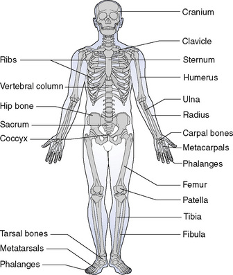
The bones of the head
For the purposes of description, the skull (Fig. 9.2) may be divided into:
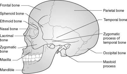
The bones of the cranium
The parietal bones form the sides and roof of the cranium; they articulate with the frontal bone, the occipital bone and with each other to form the sutures or joints of the cranium (see Chapter 11). On the internal surfaces are small grooves to carry the blood vessels supplying the brain, and the impression of the folds or convolutions of the surface of the brain can be seen. At birth there are membranous gaps in the skull at the angles of the parietal bone which are called fontanelles (see Chapter 11).
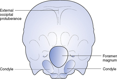
The occipital bone (Fig. 9.3) forms the back of the skull. It carries a marked prominence, the external occipital protuberance, which provides attachment for muscles. Below this there is a large oval opening, the foramen magnum, through which the cranial cavity communicates with the vertebral canal. On either side of the foramen are two smooth oval processes, the occipital condyles, for articulation with the first cervical vertebra. This joint allows the nodding movement of the head.
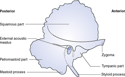
The temporal bones (Fig. 9.4) are situated at the sides and base of the skull. Each consists of four parts:
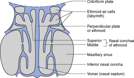
The ethmoid bone (Fig. 9.5) is very light and irregular in shape and consists of three parts:
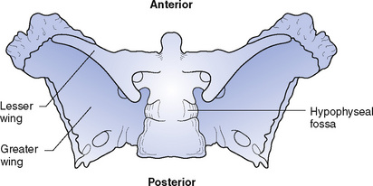
The sphenoid bone (Fig. 9.6) is situated at the base of the skull, in front of the temporal bones. It is shaped rather like a bat with outstretched wings. The body contains two large air sinuses, which communicate with the nasal cavity, and a deep depression, the hypophyseal fossa, which contains the hypophysis cerebri or pituitary gland. The greater and lesser wings are perforated by many openings for the passage of nerves and blood vessels.
< div class='tao-gold-member'>
Stay updated, free articles. Join our Telegram channel

Full access? Get Clinical Tree


