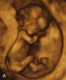
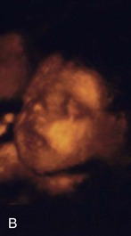
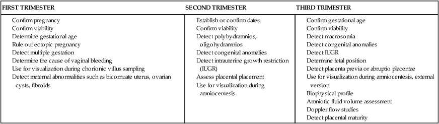
Assessment of High Risk Pregnancy


Assessment of High Risk Pregnancy


Kitty Cashion


Key Terms and Definitions


Web Resources


Additional related content can be found on the companion website at image


http://evolve.elsevier.com/Lowdermilk/Maternity/


A pproximately 500,000 of the 4 million births that occur in the United States each year are categorized as high risk because of maternal or fetal complications. A high risk pregnancy is one in which the life or health of the mother or fetus is jeopardized by a disorder coincidental with or unique to pregnancy. Care of these high risk patients requires the combined efforts of medical and nursing personnel. Factors associated with a diagnosis of a high risk pregnancy are identified in this chapter. Diagnostic techniques often used to monitor the maternal-fetal unit at risk are also described.


Assessment of Risk Factors image


Pregnancies can be designated as high risk for any of several undesirable outcomes. Those considered to be at risk for uteroplacental insufficiency (UPI), the gradual decline in delivery of needed substances by the placenta to the fetus, carry a serious threat for fetal growth restriction, intrauterine fetal death, intrapartum death, intrapartum fetal distress, and various types of neonatal morbidity.


In the past, risk factors were evaluated only from a medical standpoint; therefore only adverse medical, obstetric, or physiologic conditions were considered to place the woman at risk. Today, a more comprehensive approach to high risk pregnancy is used, and the factors associated with high risk childbearing are grouped into broad categories based on threats to health and pregnancy outcome. Categories of risk are biophysical, psychosocial, sociodemographic, and environmental (Gilbert, E.S., 2007) (Box 19-1). Risk factors are interrelated and cumulative in their effects.


BOX 19-1   Categories of High Risk Factors


Biophysical Factors


• Genetic considerations. Genetic factors may interfere with normal fetal or neonatal development, result in congenital anomalies, or create difficulties for the mother. These factors include defective genes, transmissible inherited disorders and chromosomal anomalies, multiple pregnancy, large fetal size, and ABO incompatibility.


• Nutritional status. Adequate nutrition, without which fetal growth and development cannot proceed normally, is one of the most important determinants of pregnancy outcome. Conditions that influence nutritional status include the following: young age; three pregnancies in the previous 2 years; tobacco, alcohol, or drug use; inadequate dietary intake because of chronic illness or food fads; inadequate or excessive weight gain; and hematocrit value less than 33%.


• Medical and obstetric disorders. Complications of current and past pregnancies, obstetric-related illnesses, and pregnancy losses put the patient at risk (see Box 19-2).


Psychosocial Factors


• Smoking. A strong, consistent, causal relation has been established between maternal smoking and reduced birth weight. Risks include low-birth-weight infants, higher neonatal mortality rates, increased miscarriages, and increased incidence of premature rupture of membranes. These risks are aggravated by low socioeconomic status, poor nutritional status, and concurrent use of alcohol.


• Caffeine. Birth defects in humans have not been related to caffeine consumption. However, pregnant women who consume more than 200 mg of caffeine daily (equivalent to about 12 ounces of coffee per day) may be at increased risk for miscarriage or for giving birth to infants with IUGR.


• Alcohol. Although the exact effects of alcohol in pregnancy have not been quantified and its mode of action is largely unexplained, it exerts adverse effects on the fetus, resulting in fetal alcohol syndrome, fetal alcohol effects, learning disabilities, and hyperactivity.


• Drugs. The developing fetus may be adversely affected by drugs through several mechanisms. They can be teratogenic, cause metabolic disturbances, produce chemical effects, or cause depression or alteration of CNS function. This category includes medications prescribed by a health care provider or bought over the counter, as well as commonly abused drugs such as heroin, cocaine, and marijuana. (See Chapter 20 for more information about drug and alcohol abuse.)


• Psychologic status. Childbearing triggers profound and complex physiologic, psychologic, and social changes, with evidence to suggest a relationship between emotional distress and birth complications. This risk factor includes conditions such as specific intrapsychic disturbances and addictive lifestyles; a history of child or spouse abuse; inadequate support systems; family disruption or dissolution; maternal role changes or conflicts; noncompliance with cultural norms; unsafe cultural, ethnic, or religious practices; and situational crises.


Sociodemographic Factors


• Low income. Poverty underlies many other risk factors and leads to inadequate financial resources for food and prenatal care, poor general health, increased risk of medical complications of pregnancy, and greater prevalence of adverse environmental influences.


• Lack of prenatal care. Failure to diagnose and treat complications early is a major risk factor arising from financial barriers or lack of access to care; depersonalization of the system resulting in long waits, routine visits, variability in health care personnel, and unpleasant physical surroundings; lack of understanding of the need for early and continued care or cultural beliefs that do not support the need; and fear of the health care system and its providers.


• Age. Women at both ends of the childbearing age spectrum have an increased incidence of poor outcomes; however, age may not be a risk factor in all cases. Both physiologic and psychologic risks should be evaluated.


a. Adolescents. More complications are seen in young mothers (younger than 15 years), who have a 60% higher mortality rate than those older than 20 years and in pregnancies occurring less than 6 years after menarche. Complications include anemia, preeclampsia, prolonged labor, and contracted pelvis and cephalopelvic disproportion. Long-term social implications of early motherhood are lower educational status, lower income, increased dependence on government support programs, higher divorce rates, and higher parity.


b. Mature mothers. The risks to older mothers are not from age alone but from other considerations such as number and spacing of previous pregnancies, genetic disposition of the parents, and medical history, lifestyle, nutrition, and prenatal care. The increased likelihood of chronic diseases and complications that arises from more invasive medical management of a pregnancy and labor combined with demographic characteristics put an older woman at risk. Medical conditions more likely to be experienced by mature women include hypertension and preeclampsia, diabetes, extended labor, cesarean birth, placenta previa, abruptio placentae, and death. Her fetus is at greater risk for low birth weight and macrosomia, chromosomal abnormalities, congenital malformations, and neonatal death.


• Parity. The number of previous pregnancies is a risk factor associated with age and includes all first pregnancies, especially a first pregnancy at either end of the childbearing age continuum. The incidence of preeclampsia and dystocia is increased with a first birth.


• Marital status. The increased mortality and morbidity rates for unmarried women, including an increased risk for preeclampsia, are often related to inadequate prenatal care and a young childbearing age.


• Residence. The availability and quality of prenatal care varies widely with geographic residence. Women in metropolitan areas have more prenatal visits than those in rural areas who have fewer opportunities for specialized care and consequently a higher incidence of maternal mortality. Health care in the inner city, where residents are usually poorer and begin childbearing earlier and continue for longer, may be of lower quality than in a more affluent neighborhood.


• Ethnicity. Although ethnicity by itself is not a major risk, race is an indicator of other sociodemographic risk factors. Non-Caucasian women are more than three times as likely as Caucasian women to die of pregnancy-related causes. African-American babies have the highest rates of prematurity and low birth weight, with the infant mortality rate among African-Americans being more than double that among Caucasians.


Biophysical risks include factors that originate within the mother or fetus and affect the development or functioning of either one or both. Examples include genetic disorders, nutritional and general health status, and medical or obstetric-related illnesses. Box 19-2 lists common risk factors for several pregnancy-related problems.


BOX 19-2


Specific Pregnancy Problems and Related Risk Factors


Sources: Baschat, A. A., Galan, H. L., Ross, M. G., & Gabbe, S. G. (2007). Intrauterine growth restriction. In S. Gabbe, J. Niebyl, & J. Simpson (Eds.), Obstetrics: Normal and problem pregnancies (5th ed.). Philadelphia: Churchill Livingstone; Gilbert, W. M. (2007). Amniotic fluid disorders. In S. Gabbe, J. Niebyl, & J. Simpson (Eds.), Obstetrics: Normal and problem pregnancies (5th ed.). Philadelphia: Churchill Livingstone; Resnik, R., & Creasy, R. K. (2009). Intrauterine growth restriction. In R. K. Creasy, R. Resnik, & J. D. Iams (Eds.), Creasy and Resnik’s maternal-fetal medicine: Principles and practice (6th ed.). Philadelphia: Saunders; Simpson, J. L., & Otano, L. (2007). Prenatal genetic diagnosis. In S. Gabbe, J. Niebyl, & J. Simpson (Eds.), Obstetrics: Normal and problem pregnancies (5th ed.). Philadelphia: Churchill Livingstone.


Psychosocial risks consist of maternal behaviors and adverse lifestyles that have a negative effect on the health of the mother or fetus. These risks may include emotional distress and disturbed interpersonal relationships, as well as inadequate social support and unsafe cultural practices.


Sociodemographic risks arise from the mother and her family. These risks may place the mother and fetus at risk. Examples include lack of prenatal care, low income, marital status, and ethnicity (see Box 19-1). Environmental factors include hazards in the workplace and the woman’s general environment and may include environmental chemicals (e.g., pesticides, lead, mercury), radiation, and pollutants (Silbergeld & Patrick, 2005).


Psychologic Considerations Related to High Risk Pregnancy


Once a pregnancy has been identified as high risk, the pregnant woman and her fetus will be monitored carefully throughout the remainder of the pregnancy. All women who undergo antepartal assessments are at risk for real and potential problems and may feel anxious. In most instances the tests are ordered because of suspected fetal compromise, deterioration of a maternal condition, or both. In the third trimester, pregnant women are most concerned about protecting themselves and their fetuses and consider themselves most vulnerable to outside influences. The label of high risk often increases this sense of vulnerability.


When a woman is diagnosed with a high risk pregnancy, she and her family will likely experience stress related to the diagnosis. The woman may exhibit various psychologic responses including anxiety, low self-esteem, guilt, frustration, and inability to function. A high risk pregnancy can also affect parental attachment, accomplishment of the tasks of pregnancy, and family adaptation to the pregnancy. If the woman is fearful for her own well-being, she may continue to feel ambivalence about the pregnancy or may not accept the reality of the pregnancy. She may not be able to complete preparations for the baby or go to childbirth classes if she is placed on restricted activity at home or hospitalized. The family may become frustrated because they cannot engage in activities that prepare them for parenthood. The nurse can help the woman and her family regain control and balance in their lives by providing support and encouragement, providing information about the pregnancy problem and its management, and providing opportunities to make as many choices as possible about the woman’s care.


Antepartum Testing


The major expected outcome of all antepartum testing is the detection of potential fetal compromise. Ideally, the technique used identifies fetal compromise before intrauterine asphyxia occurs so that the health care provider can take measures to prevent or minimize adverse perinatal outcomes. Antepartum testing is used primarily in patients at risk for disrupted fetal oxygenation. In most cases, monitoring begins by 32 to 34 weeks of gestation and continues regularly until birth. Assessment tests should be selected based on their effectiveness, and the results must be interpreted in light of the complete clinical picture. Box 19-3 lists common maternal and fetal indications for antepartum testing that are supported by currently available evidence (Miller, Miller, & Tucker, 2013).


The remainder of this chapter describes maternal and fetal assessment tests that are often used to monitor high risk pregnancies.


Biophysical Assessment image


Daily Fetal Movement Count


Assessment of fetal activity by the mother is a simple yet valuable method for monitoring the condition of the fetus. The daily fetal movement count (DFMC) (also called kick count) can be assessed at home and is noninvasive, inexpensive, simple to understand, and usually does not interfere with a daily routine. The DFMC is frequently used to monitor the fetus in pregnancies complicated by conditions that may affect fetal oxygenation (see Box 19-2). The presence of movements is generally a reassuring sign of fetal health.


Several different protocols are used for counting. One recommendation is to count once a day for 60 minutes. Another common recommendation is that mothers count fetal activity two or three times daily for 60 minutes each time. Except for establishing a very low number of daily fetal movements or a trend toward decreased motion, the clinical value of the absolute number of fetal movements has not been established, other than in the situation in which fetal movements cease entirely for 12 hours (the so-called fetal alarm signal). A count of fewer than three fetal movements within 1 hour warrants further evaluation by a nonstress test (NST) or contraction stress test (CST) (oxytocin challenge test [OCT]), biophysical profile (BPP), or a combination of these (see later discussion). Women should be taught the significance of the presence or absence of fetal movements (or both), the procedure for counting that is to be used, how to record findings on a daily fetal movement record, and when to notify the health care provider.


Ultrasonography


Sound is a form of wave energy that causes small particles in a medium to oscillate. The frequency of sound, which refers to the number of peaks or waves that move over a given point per unit of time, is expressed in hertz (Hz). Sound with a frequency of 1 cycle, or one peak per second, has a frequency of 1 Hz. When directional beams of sound strike an object, an echo is returned. The time delay between the emission of the sound and the return of the echo and the direction of the echo are noted. From these data the distance and location of an object can be calculated. Ultrasound is sound frequency higher than that detectable by humans (greater than 20,000 Hz). Ultrasound images are a reflection of the strength of the sending beam, the strength of the returning echo, and the density of the medium (e.g., muscle [uterus], bone, tissue [placenta], fluid, or blood) through which the beam is sent and returned.


Diagnostic ultrasonography is an important, safe technique in antepartum fetal surveillance. It provides critical information to health care providers regarding fetal activity and gestational age, normal versus abnormal fetal growth curves, visual assistance with which invasive tests may be performed more safely, fetal and placental anatomy, and fetal well-being (Richards, 2007). Ultrasound examination can be performed abdominally or transvaginally during pregnancy. Both methods produce a two- or three-dimensional view from which a pictorial image is obtained (Fig. 19-1, A, B). It is also possible to produce a four-dimensional image. Abdominal ultrasonography is more useful after the first trimester when the pregnant uterus becomes an abdominal organ. During the procedure, the woman usually should have a full bladder to displace the uterus upward to provide a better image of the fetus. Transmission gel or paste is applied to the woman’s abdomen before a transducer is moved over the skin to enhance transmission and reception of the sound waves. She is positioned with small pillows under her head and knees. The display panel is positioned so that the woman or her partner (or both) can observe the images on the screen if they desire.


Transvaginal ultrasonography, in which the probe is inserted into the vagina, allows pelvic anatomic features to be evaluated in greater detail and intrauterine pregnancy to be diagnosed earlier. A transvaginal ultrasound examination is well tolerated by most pregnant women because it removes the need for a full bladder. It is especially useful in obese women whose thick abdominal layers cannot be penetrated adequately with an abdominal approach. A transvaginal ultrasound may be performed with the woman in a lithotomy position or with her pelvis elevated by towels, cushions or a folded pillow. This pelvic tilt is optimal to image the pelvic structures. A protective cover such as a condom, the finger of a clean rubber surgical glove, or a special probe cover provided by the manufacturer is used to cover the transducer probe. The probe is lubricated with a water-soluble gel and placed in the vagina either by the examiner or by the woman herself. During the examination the position of the probe or the tilt of the examining table may be changed so that the complete pelvis is in view. The procedure is not physically painful, although the woman will feel pressure as the probe is moved. Transvaginal ultrasonography is optimally used in the first trimester to detect ectopic pregnancies, monitor the developing embryo, help identify abnormalities, and help establish gestational age. In some instances, it may be used as an adjunct to abdominal scanning to evaluate preterm labor in second- and third-trimester pregnancies.


Levels of ultrasonography


The American College of Obstetricians and Gynecologists (ACOG) (2004) describes three levels of ultrasonography. The standard examination is used most frequently and can be performed by ultrasonographers or other heath care professionals, including nurses, who have had special training. Indications for standard ultrasonography are described in detail in the next section. Its primary uses are to detect fetal viability, determine the presentation of the fetus, assess gestational age, locate the placenta, examine the fetal anatomic structures for malformations, and determine amniotic fluid volume (AFV). Limited examinations are performed for specific indications such as identifying fetal presentation during labor or evaluating fetal heart rate (FHR) activity when it is not detected by other methods (ACOG). Specialized or targeted examinations are performed if a woman is suspected of carrying an anatomically or a physiologically abnormal fetus. Indications for this comprehensive examination include abnormal findings on clinical examination, especially with polyhydramnios or oligohydramnios, elevated alpha-fetoprotein (AFP) levels, and a history of offspring with anomalies that can be detected by ultrasound examination. Specialized ultrasonography is performed by highly trained and experienced personnel (ACOG).


Indications for use


Major indications for obstetric sonography are listed by trimester in Table 19-1. During the first trimester, ultrasound examination is performed to obtain information regarding the number, size, and location of gestational sacs; the presence or absence of fetal cardiac and body movements; the presence or absence of uterine abnormalities (e.g., bicornuate uterus or fibroids) or adnexal masses (e.g., ovarian cysts or an ectopic pregnancy); and pregnancy dating (by measuring the crown-rump length).


During the second and third trimesters, information regarding the following conditions is sought: fetal viability, number, position, gestational age, growth pattern, and anomalies; AFV; placental location and maturity; presence of uterine fibroids or anomalies; presence of adnexal masses; and cervical length.


Ultrasonography provides earlier diagnoses, allowing therapy to be instituted sooner in the pregnancy, thereby decreasing the severity and duration of morbidity, both physical and emotional, for the family. For instance, early diagnosis of a fetal anomaly gives the family choices such as intrauterine surgery or other therapy for the fetus, termination of the pregnancy, or preparation for the care of an infant with a disorder.


Stay updated, free articles. Join our Telegram channel

Oct 8, 2016 | Posted by in NURSING | Comments Off on Assessment of High Risk Pregnancy

Full access? Get Clinical Tree

Get Clinical Tree app for offline access