CHAPTER 3
Anatomy and Physiology of the Vascular System
Diane Treat-Jacobson
OBJECTIVES
1. Describe the anatomy and physiology of the arterial, venous, and lymphatic systems.
2. Describe the changes that occur in the vascular system with aging.
Introduction/Overview
This chapter reviews the structure and function of the three major components of the vascular system: the arterial, venous, and lymphatic systems. The purpose of the vascular system is to transport blood from the heart to all parts of the body, to allow for the exchange of substances between the blood and the tissues, and to return blood to the heart. The vessel size, wall thickness, and internal pressure within vessels vary depending on vessel type and function.
 I. Arterial Circulation (Graham & Ford, 1999; Guyton & Hall, 2000; Spence, 1982; Tottora & Grabowski, 2003)
A. General Arterial Anatomy and Physiology: Conductive versus Distributive Vessels
1. Conductive vessels are relatively straight with few branches
2. Distributive vessels arise from conductive arteries and have multiple branches
3. Arteries, arterioles, capillaries
a. Arteries transport blood to the tissues under high pressure (mean pressure 100 mm Hg)
b. Arterioles: the last small branch of the arterial system; controls flow into the capillary. Proximal pressure approximately 85 mm Hg and distal pressure approximately 30 mm Hg
c. Capillary: fluid, nutrients, electrolytes, and hormones are exchanged between the blood and the interstitial fluid. Arterial end capillary pressure approximately 30 mm Hg; venous end capillary pressure approximately 10 mm Hg

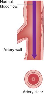
FIGURE 3.1 Cross-section of arterial wall.
B. Arterial Structure (see Fig. 3-1)
1. Intima
a. The innermost layer, lining the lumen of the arterial wall
b. Consists of a single layer of endothelial cells
c. Separated from the media by the internal elastic membrane
d. Nutrients obtained by diffusion from the arterial lumen
2. Media
a. Thick layer of smooth muscle cells, collagen, and elastic fibers
b. Separated from the adventitia by the external elastic membrane
c. Inner portion receives nutrients by diffusion from the arterial lumen; outer portion receives nutrients from small blood vessels that penetrate the arterial wall
3. Adventitia
a. Outer layer of the artery
b. Composed of connective tissue, collagen, and elastic fibers
c. Provides majority of strength of the arterial wall
d. Nutrients obtained from small blood vessels that penetrate the arterial wall
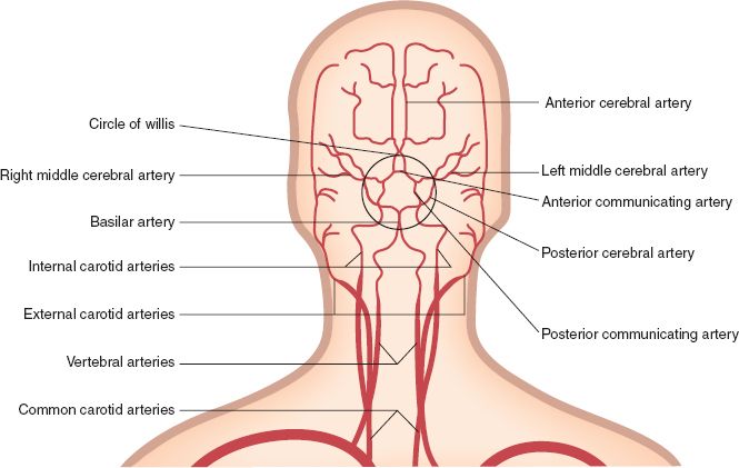
C. Arteries of the Head and Neck (see Fig. 3-2)
1. After arising from the aortic arch, the brachiocephalic artery divides into the right subclavian artery and the right common carotid artery
2. Right and left common carotid arteries supply most of the blood to the head and neck
3. Common carotid arteries bifurcate as follows:
a. Carotid sinus: formed at the point of bifurcation
b. Right and left external carotid arteries
1) Supply most of head and neck except for the brain
2) Lie more superficially than the internal carotid arteries
3) Terminate as the maxillary and superficial temporal arteries supplying muscles of mastication, the nose, pharynx, palate, teeth, and some neck muscles
4) Give off the occipital artery to the posterior region of the scalp

FIGURE 3.2 Arteries of the head and neck.
5) Send branches to supply the thyroid gland, tongue, skin, and muscles of the face
c. Right and left internal carotid arteries
1) Enter the cranial cavity through the carotid canal of the temporal bone
2) Supply the brain through the terminal branches: the anterior cerebral artery, the middle cerebral artery
3) Right and left internal carotid arteries are connected by the anterior communicating artery
d. Vertebral artery
1) First branch of the subclavian artery
2) Reaches the cranial cavity by passing through the transverse foramen of the cervical vertebrae and the foramen magnum
3) Right and left vertebral arteries join to form the basilar artery
4) Basilar artery supplies pons and cerebellum
5) Basilar artery terminates as the right and left posterior cerebral arteries
e. Circle of Willis
1) Anterior communicating artery, anterior cerebral artery, posterior communicating artery, and posterior cerebral artery join to form the Circle of Willis
2) Provides the potential for collateral flow to compensate for the loss of a carotid or vertebral artery
D. Arteries of the Upper Extremities
1. Supplied by the subclavian arteries
2. First branches off the subclavian artery do not supply the upper limbs
a. Thyrocervical trunk sends branches to lower neck and dorsal scapular regions
b. Costocervical trunk sends branches to the intercostal region
c. Internal thoracic (internal mammary artery) supplies branches to the thoracic and intercostal muscles
3. Subclavian artery becomes the axillary artery after passing through the clavicle
a. Branches to the lateral wall of the thorax
1) Thoracocranial trunk
2) Lateral thoracic artery
3) Subscapular artery
4) Thoracodorsal artery
b. Branches to region around proximal end of the humerus (circumflex arteries)
c. Becomes brachial artery, traveling down medial side of humerus and supplying muscles of the arm
d. Brachial artery branches as follows:
1) Ulnar artery down anterior surface of forearm along ulna
2) Radial artery down anterior surface of forearm along radius
3) Ulnar and radial arteries join in the hand through interconnecting branches of the superficial and deep palmar arteries
4) Digital arteries arise from the palmar arches and supply the fingers
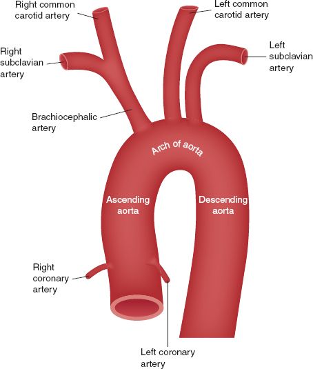
E. Aorta (see Fig. 3-3)
1. Ascending aorta
a. Passes in front of the pulmonary artery
b. Branches include right and left coronary arteries

FIGURE 3.3 Aortic arch.
2. Aortic arch
a. Aorta arches dorsally as it leaves the pericardial sac
b. Branches include brachiocephalic artery, left common carotid artery, and left subclavian artery
c. Furnishes all blood to head, neck, and upper limbs
3. Thoracic aorta gives rise to the following:
a. Posterior intercostal arteries
b. Bronchial artery
c. Esophageal artery
d. Superior phrenic artery
F. Abdominal Aorta/Mesenteric Arteries
1. Lumbar arteries supply posterior abdominal walls
2. Inferior phrenic arteries supply underside of diaphragm
3. Celiac artery
a. Arises just below inferior phrenic arteries
b. Divides as follows:
1) Left gastric artery supplies lower part of esophagus and part of the stomach
2) Splenic artery
a) Supplies the spleen
b) Gives branches to the pancreas and stomach
c) Branches to the left gastroepiploic artery supplies greater curvature of the stomach
3) Hepatic artery
a) Supplies liver
b) Sends branches to the stomach, duodenum, pancreas, and gallbladder
4. Superior mesenteric artery
a. Arises below celiac artery
b. Supplies all of small intestine, cecum, and the ascending and transverse colon of the large intestine
5. Suprarenal arteries
a. Branch from the aorta at the level of the superior mesenteric artery
b. Supply adrenal glands
6. Renal arteries
a. Arise from aorta inferior to superior mesenteric artery
b. Supply the kidneys
7. Ovarian arteries (women)
a. Arise from aorta below renal arteries
b. Supply ovaries
c. Send branches to the ureters and uterine tubes
8. Testicular (internal spermatic) arteries (men)
a. Arise from aorta below renal arteries
b. Pass through inguinal canal to the scrotum; supply the testes
9. Inferior mesenteric artery
a. Final branch of abdominal aorta
b. Supplies part of transverse colon, descending colon, sigmoid colon, and rectum

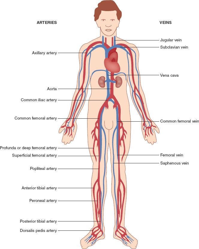
FIGURE 3.4 Arterial and venous anatomy.


