9: Diabetes mellitus

Chapter 9 Diabetes mellitus


RELEVANT ANATOMY AND PHYSIOLOGY OF PANCREAS


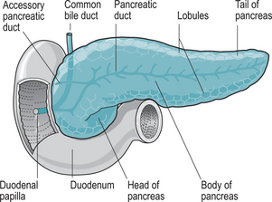
The pancreas is an oblong gland, approximately 12.5 cm long and 2.5 cm thick. It is situated in the epigastric region posterior to the greater curvature of the stomach, and is connected by two ducts to the duodenum (Fig. 9.1). The head is the expanded portion situated near the ‘C’ shaped curve of the duodenum, superior and to the left are the central body and tail.


The pancreas is both an endocrine and an exocrine gland. Endocrine glands secrete hormones into the blood, while exocrine glands secrete their products through ducts into body cavities or onto body surfaces. The largest portion of the pancreatic cell mass is composed of acini – clusters of cells which constitute the exocrine or digestive portion of the gland, secreting a mixture of digestive fluid and enzymes called pancreatic juice into the duct system. Pancreatic secretions pass from the secreting cells into ducts that ultimately convey the secretions into the small intestine.


Pancreatic juice is a clear, colourless fluid containing water, salts, sodium bicarbonate and enzymes which digest carbohydrate, fat and protein. The main enzymes involved in this process are amylase, lipase and trypsin. Normally, the amount secreted is approximately 1200–1500 mL/day in the adult.


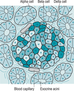
Interspersed within the acini are small clusters of cells called the islets of Langerhans or pancreatic islets, which form the endocrine portion of the pancreas (Fig. 9.2). There are four types of cells within the islets, which secrete the following hormones:


A brief synopsis of the endocrine function of the pancreas is provided to emphasize the importance of the pancreatic function in metabolic homeostasis.


Pancreatic polypeptide regulates the release of pancreatic digestive hormones. Somatostatin, or growth hormone inhibiting factor (GHIF), acts as a paracrine (a local hormone which acts on neighbouring cells), to inhibit the secretion of insulin and glucagon. Glucagon and insulin are the endocrine secretions of the pancreas and are concerned with the regulation of blood glucose.


If glucose is not required by the body immediately for energy, then it is converted to glycogen and stored in the liver and skeletal muscle. Glucagon acts mainly on the liver, and its principal activity is to increase the blood glucose level by accelerating the conversion of glycogen to glucose. This process is known as glycogenolysis. Glucagon also aids the conversion of other nutrients such as certain amino acids and lactic acid into glucose, known as gluconeogenesis. The liver then releases glucose into the bloodstream and the blood glucose level rises.


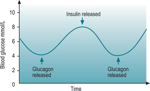
Secretion of glucagon is via a negative feedback system (Fig. 9.3); if the blood glucose level falls below normal, chemical sensors stimulate the alpha cells to secrete glucagon. When blood glucose level rises, the cells are no longer stimulated and production ceases. Exercise and protein-heavy meals also stimulate glucagon secretion. Somatostatin inhibits glucagon secretion.


Emotional stress commonly raises blood glucose levels. Cortisol is a corticosteroid hormone produced by the adrenal cortex and is involved in the response to stress. It increases blood pressure and blood glucose levels. Cortisol has widespread actions which help to restore homeostasis after stress. Cortisol acts as a physiological antagonist to insulin by promoting gluconeogenesis, the breakdown of fats and proteins and the mobilization of amino acids and ketone bodies. This leads to an increase in blood glucose levels and the storage of glycogen in the liver (Marieb & Hoehn 2007).


Insulin is produced by beta cells. Its physiological action is the reverse of glucagon. Insulin accelerates the transport of glucose from the blood into the cells, especially skeletal muscle cells, and thereby decreases the blood glucose level. Insulin also accelerates the conversion of glucose to glycogen. This is known as glycogenesis. It decreases glycogenolysis and gluconeogenesis and stimulates the conversion of glucose and other nutrients into fatty acids (lipogenesis). Insulin also stimulates protein synthesis.


Regulation of insulin is determined by the glucose level in the blood. Increased blood glucose levels stimulate insulin secretion, while decreased blood glucose levels inhibit it (Fig. 9.3). Increased levels of certain amino acids also stimulate insulin release. Human growth hormone (hGH) and adrenocorticotrophic hormone (ACTH) both raise blood glucose levels, and the rise in blood glucose level stimulates insulin secretion. Somatostatin inhibits the secretion of insulin.


Metabolism


The word ‘metabolism’ is translated from ‘metabolismos’, the Greek word for ‘change’, or ‘overthrow’. Metabolism is the biochemical modification of chemical compounds in living organisms and cells. This includes the biosynthesis of complex organic molecules and their breakdown. Nutrients are digested and broken down into chemical compounds; some of these chemical compounds recombine to form other compounds or building blocks (anabolism) and some chemicals break down to provide energy (catabolism).


Metabolism usually consists of sequences of enzymatic steps, or metabolic pathways which are a series of chemical reactions occurring within a cell, catalyzed by enzymes. Many of these pathways are elaborate, and involve a step by step modification of the initial substance to shape it into the product with the exact chemical structure desired. Catabolic pathways break down complex molecules into simple compounds, while anabolic pathways create building blocks and compounds from simple precursors.


TYPES OF DIABETES MELLITUS


There are two main types of diabetes mellitus. Type 1, which was formerly known as insulin dependent diabetes mellitus (IDDM), occurs mainly in the young. Type 2, which was known as non-insulin dependent diabetes mellitus (NIDDM) was said to occur in the middle-aged, overweight adult. These are rather simplistic generalizations and not entirely accurate, as insulin may be required in individuals with type 2 diabetes mellitus and also type 2 diabetes is now occurring in younger age groups. Other forms of diabetes include impaired glucose regulation, which refers to a metabolic state between normal glucose homeostasis and diabetes. This condition may be a risk factor for the development of diabetes in the future. Maturity onset diabetes of the young (MODY) is a rare type of diabetes associated with genetic defects. For the midwife, this discussion will focus on the two main types of diabetes mellitus and the form of diabetes which occurs in pregnancy – gestational diabetes mellitus.


Type 1 diabetes mellitus


Aetiology


The aetiology of type 1 diabetes mellitus is thought to be multifactorial and far from being completely understood. It is considered to have an autoimmune basis and a number of risk factors have been identified, which may be divided into genetic and environmental. These risk factors include links to chromosomes and several chromosomal regions have been implicated. The most significant gene loci defining the risk of type 1 diabetes are to be found within leucocyte antigen (HLA) gene region (BMA 2004).


There has been a rapidly rising incidence in migrant populations suggesting environmental links, for example viral infections. It has been observed that IgM antibodies to Coxsackie B virus are found in 25–35% of newly diagnosed diabetics (Kanno et al 2006). An increased incidence of type 1 diabetes has been noted in individuals with congenital rubella, and cytomegalovirus DNA has also been found in 22% of newly diagnosed diabetics (Jaeckel et al 2002).


It has also been suggested that those with diabetes were breast-fed for a significantly shorter time than non-diabetic infants, and that the incidence of diabetes can be modified by the exclusion of cow’s milk, suggesting that early exposure to cow’s milk may be an important factor in the aetiology of type 1 diabetes (BMA 2004). The condition varies considerably in incidence between cultures with the 10 countries with the highest proportion of sufferers being India, China, USA, Indonesia, Japan, Pakistan, Russia, Brazil, Italy and Bangladesh (WHO 2006).


Pathophysiology


Type 1 diabetes mellitus occurs as the result of rapid and progressive loss of beta cells, resulting in an absolute deficiency of insulin, and is thought to be precipitated by a response to physical or psychological stress.


Lack of insulin has a domino effect, as cells cannot therefore obtain glucose for metabolism and this illustrates how the clinical features arise. Ingested glucose which is unable to enter cells accumulates in the blood leading to hyperglycaemia; this high blood glucose level circulates throughout the body including the renal system, which cannot cope with the increasing amounts of glucose. As a consequence, the renal threshold is exceeded, and glucose appears in the urine, resulting in glycosuria. Glucose is highly osmotic (it attracts and holds water to itself), therefore, as glucose is lost in the urine, large volumes of water are lost with it. This is known as polyuria. Furthermore, nocturia may occur resulting in disturbed sleep patterns. As fluid loss continues, symptoms of dehydration occur, resulting in copious drinking in response to severe thirst – polydipsia.


As the cell requirements for glucose are not being met, the response is for increased secretion of glucagon. Glycogen is converted back into glucose and released by the liver – glycogenolysis. However, glycogen storage is quickly depleted and in an attempt to supply the cells there is a breakdown of body fat and protein to produce glucose – gluconeogenesis.


Body cells are not being supplied with glucose, so may stimulate appetite – polyphagia, although this may be tempered somewhat because of nausea and/or vomiting. Weight loss occurs due to depletion of body fat and insulin stores, which together with cell deprivation of glucose, fluids and electrolyte imbalance results in muscle weakness and exhaustion. This exhaustion may be so overwhelming as to mask the presence of the other clinical features in the affected individual.


Because there is no insulin, fat metabolism results in ketone bodies being produced. These accumulate in the blood and urine, causing ketonaemia and ketonuria. Ketone bodies are weak acids, which release free hydrogen ions causing metabolic acidosis. Excess acidity in the blood causes hyperventilation – the body’s response to removing excessive hydrogen ions.


Uncontrolled and untreated, these features will lead to a life-threatening condition known as diabetic ketoacidosis (McIntyre & Strachan 2000).


Management


The mainstay of care is dietary advice, insulin, blood glucose monitoring, education and psychological care. At the time of diagnosis, intensive educational and psychological support is provided and as the patient learns and becomes more familiar with the condition, she will be encouraged to manage her own condition. Newly diagnosed individuals may experience a wide range of emotions including shock, anger and fear. Because of this, education should be given in small bites and verbal information should be backed up with written reinforcement. Information on how to contact the diabetic liaison nurse by telephone or a local diabetic self-help group is also helpful.


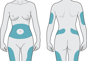
Giving insulin injections is usually a concern, and must be demonstrated and practised carefully by the individual until they feel confident. Alcohol swabbing is contraindicated, as it causes the skin to toughen, making injections more difficult. Needle length, angle of injection, as well as suitable sites for injection must also be addressed (Box 9.1, Fig. 9.4). Various devices are available to deliver insulin to the individual, including insulin pens, which are loaded with a single use cartridge containing the required dose.


Box 9.1 Administration of insulin


Insulin is available in different presentations depending on the device used. Insulin vials are used with insulin syringes. Insulin cartridges are used with reusable pens and disposable pre-filled pens are also available.


Insulin is administered by subcutaneous injection. There are three main areas for injection – abdomen, buttocks and thighs (Fig. 9.4). The upper arm is the least preferred site as it has little subcutaneous tissue. Overuse of one site can lead to fat hypertrophy and loss of sensitivity. If insulin is injected into fatty tissue, this will result in slower and more erratic absorption of insulin, resulting in variable and unstable blood glucose readings. It is advisable to use a different site each day and by rotating sites, this ensures the insulin will act quickly and effectively.


To reduce the discomfort of an injection, some newly diagnosed diabetics hold an ice cube against the skin for a few seconds, just prior to injection. This can help numb the area to any pain.


On-going education is provided regarding the type of insulin used, how it works, and how to adjust the dose according to the patient’s level of activity and blood glucose levels.


Capillary blood glucose monitoring is also demonstrated and practised until the individual feels proficient. Most newly diagnosed individuals choose to purchase a blood glucose meter (not available on prescription, although the test strips and lancets for use with the meter are available on prescription). Some diabetic nursing teams can supply glucose meters free of charge.


Glucose tests are usually carried out four times daily before meals and at bedtime and should aim to keep blood glucose levels between 4–7 mmol/L. The advantage of daily blood glucose monitoring is that it provides day-to-day control and greater patient involvement in managing their diabetes.


Glycosylated haemoglobin (HbA1c) is a valuable blood test which identifies the proportion of haemoglobin bound to glucose, and averages blood glucose levels over a 6–8 week period – the life span of a red blood cell – thereby providing an estimation of diabetic glycaemic control over this time. Ideally, the target of glycosylated haemoglobin is ≤7% for people with type 1 diabetes and between 6.5% and 7.5% for people with type 2 diabetes.


Urinalysis is not routinely carried out, as it is unhelpful for all those using insulin. It does not diagnose low blood glucose levels nor can it be used to identify a hypoglycaemic event. Furthermore, it is considered that urinalysis provides neither an accurate nor a precise measurement of high or low blood glucose levels (Diabetes UK 2006).


First aid dietary advice may be given by the nurse but mainly the dietician assesses the patient and provides individualized education. A healthy diet, which includes high levels of complex carbohydrate and fibre, moderate protein, and low fat, particularly saturated fats, is recommended. Regular meals, five portions of fruit and vegetables, plenty of water, avoiding sugary drinks and cakes and reducing salt intake will go far to ensure a healthy diet.


The most important issues are that the individual must be supported in this very difficult time and that she has access to specialist diabetic care resources and self-help groups. ‘Buddy’ support groups where an experienced diabetic teams up with a newly diagnosed individual are useful but must not replace professional support (Scottish Diabetes Framework 2006).


< div class='tao-gold-member'>

Stay updated, free articles. Join our Telegram channel

Jul 11, 2016 | Posted by in MIDWIFERY | Comments Off on 9: Diabetes mellitus

Full access? Get Clinical Tree

Get Clinical Tree app for offline access