DIABETIC KETOACIDOSIS (DKA)
I. Definition
A. A state of intracellular dehydration that results from elevated blood glucose levels
B. Hyperglycemia increases serum osmolality, causing a shift of intracellular water into the intravascular space.
C. In addition to hyperglycemia, DKA is characterized by hyperketonemia and an acidotic pH.
II. Incidence/predisposing factors
A. Accounts for approximately 14% of all hospital admissions among diabetic patients; although it usually occurs in those with type 1 diabetes, it may occur in patient with type 2 diabetes as well.
B. Occurs in approximately 46 of 10,000 diabetic patients
C. Increasing incidence among patients with insulin pumps
D. Mortality rate is approximately 5%.
E. Poor patient compliance—Common causes include the following:
1. Lack or omission of insulin; classically, the patient stops taking or fails to appropriate insulin
2. Too much food or insufficient exercise without an appropriate amount of insulin
3. Failure to consume extra fluids and insulin during illness or acute stress
F. Pancreatitis
G. Sepsis/infection
H. Surgery or trauma in the patient with diabetes mellitus
IV. Laboratory/diagnostic findings
A. Serum glucose levels greater than 250 mg/dl and frequently greater than 300 mg/dl
B. Arterial pH usually less than 7.3 and PCO2 (partial pressure of CO2) less than 40 mmHg, indicating metabolic acidosis; HCO3 less than 15 mEq/L
C. Ketones present in serum and urine
D. Hyperkalemia is related to shifting of hydrogen ions intracellularly in an attempt to buffer the acidosis; subsequently, hydrogen ions are exchanged for potassium ions.
E. Increased BUN level related to dehydration
F. Glycosuria
G. Increased hematocrit level related to dehydration
H. Leukocytosis—WBC count may be 25,000/microliter.
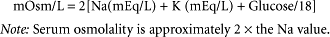
I. Serum hyperosmolality (greater than 280 mOsm/L) is common. Note: Osmolality greater than 320 to 330 mOsm/L usually results in coma. To effectively measure serum osmolality, use the following equation:
J. Expect an increased anion gap = Na − (HCO3 + Cl)
1. Normal anion gap is 7 to 17 mEq/L.
2. Note that the higher the anion gap is, the higher is the patient’s acuity.
K. Hypercholesterolemia may be present.
L. Hypertriglyceridemia may be present.
M. Hyperamylasemia may be present.
V. Management


A. Critical care monitoring is indicated. Consider invasive monitoring (e.g., central venous pressure/pulmonary arterial catheter) based on the patient’s history of cardiovascular and/or pulmonary disease (e.g., congestive heart failure, pulmonary edema).
B. Parenteral fluid replacement should be initiated with 0.9% normal saline (NS) at 1000 ml/hour for 1 to 2 hours; this should be followed by administration of 300 to 500 ml/hour for 4 hours to correct a usual fluid deficit of 4 to 8 L.
1. Once dehydration improves, 250 ml/hour is recommended.
2. Expect to order approximately 4 to 8 L fluid to be administered during the first 24 hours of treatment.
C. Potassium values should be closely monitored during fluid resuscitation.
D. Isotonic fluids are generally used until the patient is hemodynamically stable.
E. Once the patient is hemodynamically stable, hypotonic solutions (e.g., ½ NS) are used to promote intracellular hydration.
F. As the patient’s glucose levels fall to approximately 250 mg/dl, IV fluids are changed to dextrose-containing agents such as D5½ NS to prevent hypoglycemia and cerebral edema caused by lowering glucose too rapidly.
Stay updated, free articles. Join our Telegram channel
Full access? Get Clinical Tree
Get Clinical Tree app for offline access
