Head trauma/traumatic brain injury, 959.01
Spinal cord trauma, 952.9
I. Head trauma accounts for two thirds of all casualties of motor vehicle accidents.
A. Head trauma is the leading cause of death in all trauma cases.
B. Anatomic structures and physiologic functions of the head provide protection for the brain.
1. Scalp
2. Skull
3. Cerebral meninges (PAD—pia mater, arachnoid, dura mater)
4. CSF
C. The brain is dependent on glucose (25%) and oxygen (20%) for functioning.
II. Mechanism of injury
A. Acceleration
B. Deceleration
C. Deformation
III. Type of injury
A. Blunt trauma
B. Penetrating injury
1. High-velocity object
2. Low-velocity object
C. Coup-contrecoup injuries: brain tissue injury directly at the site of impact (coup) and at the pole opposite the site of impact (contrecoup) that may be caused by movement of cranial contents within the skull
V. Clinical manifestations
A. Many have been discussed previously under specific types of head injuries.
B. When the patient is beginning to decompensate, symptoms of Cushing’s triad may develop.
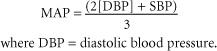
CPP = MAP − ICP
where MAP = mean arterial pressure and ICP = intracranial pressure.
2. Decreased respiratory rate
3. Decreased heart rate
C. Neurologic examination
1. AVPU—A = awake, V = responds to verbal stimuli, P = responds to painful stimuli, U = unresponsive
2. Glasgow coma scale (Individuals with subdural hematoma and a score less than or equal to 8 have a poor prognosis.)
3. Posturing
a. Decorticate: flexion of upper extremities with extension of lower extremities
b. Decerebrate: extension of upper extremities and lower extremities (More of the brain stem is involved.)
D. Laboratory findings in patients with diabetes insipidus
1. Electrolytes: increased sodium, decreased potassium, and so forth
2. Increased serum osmolality (greater than 275-285 mOsm/kg)
3. Decreased urine osmolality (N = 300-900 mOsm/kg)
4. Decreased urine specific gravity (N = 1.010-1.030)
5. CBC; hemoconcentration (hematocrit and hemoglobin may be falsely high)
VI. Management
A. Consult a neurosurgeon.
B. Prevent hypotension and hypoxemia.
1. According to Brain Trauma Foundation guidelines, resuscitation should be aimed at maintaining a systolic blood pressure (SBP) greater than 90 mmHg and a partial pressure of arterial oxygen (PaO2) greater than 60 mmHg.
2. Attempt to maintain an MAP greater than 90 mmHg.
3. If the patient is hypotensive, administer 1-2 L of isotonic crystalloid solution immediately. Avoid overhydration as attempts are made to restore adequate blood pressure (MAP greater than 90 mmHg).
4. The literature has increasingly recommended small-volume resuscitation with hypertonic/hyperosmolar solutions (e.g., hypertonic saline, dextran) because of the findings that these solutions enhance intracranial compliance, resulting in a minimal increase in ICP.
C. Hyperventilation/hyperoxia
1. Hyperventilation (PaCO2 [partial pressure of carbon dioxide in arterial blood], 25-30 mmHg) has been used for decades to cause cerebral vasoconstriction and thereby lower ICP. Cerebral vasoconstriction caused by hyperventilation has also been known to cause cerebral ischemia.
2. Experts now recommend that hyperventilation should not be used routinely, especially during the first 24 hours after injury, unless ICP is severely high or the patient requires suctioning.
a. Brain Trauma Foundation guidelines specifically recommend that hyperventilation to bring PaCO2 to less than 35 mmHg should be undertaken only if a measured increase in ICP is reported, or if increased ICP is suspected because of physical signs, while intracranial hypertension is refractory to other interventions.
b. Other methods known to control ICP (e.g., elevating the head of the bed, sedation, paralysis, mannitol, CSF drainage) should be instituted first.
3. Other studies support that hyperoxia during acute hyperventilation improves oxygen delivery to the brain, as indicated by improved jugular venous bulb saturation and arteriovenous oxygen content difference. Moderate hyperventilation to a PaCO2 of 30 mmHg in combination with a higher PaO2 may be beneficial in patients with head injury.
4. Nasal intubation and a nasogastric (NG) tube have traditionally been preferred over oral intubation, given the possibility of cervical spine injury, with the exception of basilar skull fracture.
D. Central nervous system (CNS) depressants
1. Opioid sedatives help lower ICP by reducing metabolic demand and relieving anxiety and pain. Naloxone (Narcan) can reverse the effects of a narcotic sedative when a neurologic assessment is conducted.
2. Short-acting opioids are the best choices.
a. IV fentanyl, 2-20 mcg/kg over 1 to 2 minutes, up to 50 mcg/kg, or sufentanil (Sufenta), 1-8 mcg/kg/minute
b. These may be supplemented with propofol (Diprivan) or benzodiazepines (diazepam [Valium] or lorazepam [Ativan]) if the patient remains agitated or ICP remains elevated.
3. Neuromuscular blocking agents
a. Vecuronium (Norcuron, 0.04-0.1 mg/kg IV) or doxacurium (Nuromax, 0.05 mg/kg)
b. Can be used to help lower ICP in patients in whom confusion, posturing, or severe agitation is interfering with treatment or diagnostic testing
c. Patient must be sedated and intubated with an adequate set rate on the ventilator.
E. Steroids
1. Corticosteroids remain controversial.
a. Theoretically, these drugs work by preventing fluid from entering the cells and by increasing blood vessel diameter, thereby promoting cerebral blood flow.
b. However, according to Brain Trauma Foundation guidelines, the use of steroids is not recommended for improving outcomes or reducing ICP in patients with severe head injury.
2. Many practitioners believe that the risks of steroids (e.g., compromise of immune function, infection, delayed wound healing, gastric distress) far outweigh the benefits.
a. No significant improvement or reduction in ICP has been noted with the use of steroids.
b. A study performed on rats found that low doses of dexamethasone (Decadron) appear to be beneficial for the treatment of cerebral trauma because they decrease brain water content.
F. Osmotherapy (mannitol)
1. Very useful in certain patients; drug of choice in an emergency situation when brain herniation is pending
2. Mannitol creates an osmotic gradient across the blood–brain barrier that pulls water from the CNS into the intravascular space.
3. Mannitol may enhance cerebral oxygen delivery via decreased blood viscosity, increased CPP, or both.
4. Mannitol may also provide some cytoprotective effects through oxygen-free radical scavenging.
5. Administer as a bolus (0.25-1 g/kg).
6. Results are quick, occurring usually within 10 to 20 minutes, and can last up to 6 hours.
7. Monitor serum osmolarity, and maintain at less than 320 mOsm. Monitor electrolytes as well, and replace as needed.
8. Monitor blood pressure closely.
a. Hypotension is an adverse effect that results from dehydration caused by diuresis.
b. The goal is to attain beneficial effects without inducing dehydration.
9. Volume replacement may be necessary to keep the patient euvolemic and to prevent hypotension (e.g., replace urinary output volume-for-volume each hour, or one-half volume-for-volume each hour, with isotonic crystalloids).
10. An indwelling urinary catheter should be in place and urine output recorded each hour.
11. Some investigators are replacing mannitol with loop diuretics such as furosemide (Lasix) and ethacrynic acid (Edecrin) to control ICP, believing that these drugs are less likely to cause severe dehydration.
H. It is recommended that anticonvulsants (phenytoin [Dilantin], carbamazepine [Mazapine]) may be used to prevent early posttraumatic seizures in those at high risk after a head injury. Prophylactic use is not recommended for preventing late posttraumatic seizures.
I. Patient must undergo ongoing neurologic assessment for evaluation of treatment effectiveness and the need for additional interventions.
1. Evaluation of the Glasgow coma scale score may be necessary every 30 to 60 minutes for the first 24 hours. Note whether the patient is receiving sedation and/or paralytics because these may lower the score.
2. Pupil size and reaction
3. Vital signs
J. It is suggested that by the seventh day after injury, 140% of resting metabolism expenditure is replaced in nonparalyzed patients, and that 100% of resting metabolism expenditure occurs in paralyzed patients who use enteral or parenteral formulas that contain at least 15% of calories as protein.
K. All patients with head injury are presumed to have a cervical spine injury until proven otherwise. Once the patient’s condition has stabilized, a cervical spine series should be ordered.
L. Avoid any condition (e.g., fever, pain, shivering) that increases metabolic rate and therefore increases the demand for O2 and glucose.
M. Prospective treatment
Get Clinical Tree app for offline access

1. Polyethylene glycol superoxide dismutase is a drug that investigators are exploring to determine whether it can decrease toxicity to the brain associated with free radicals that are released during trauma. This drug appears to improve clinical outcomes in head trauma victims.
2. Lazaroids—synthetic nonglucocorticoids—constitute a group of experimental drugs that may slow cellular membrane breakdown after head injury by inhibiting lipid peroxidation.
4. Hypothermia had been thought to possibly improve clinical outcomes through control of ICP.
a. Lowering the temperature to 89.6° F to 91.4° F has resulted in improved outcomes in animals, and the procedure has been tested in humans.
b. Recent studies found no evidence that hypothermia is beneficial in the treatment of patients with head injury.
5. Decompressive craniectomy has been recognized over the past few years for its use in patients with malignant ICP caused by traumatic brain injury (TBI).


a. Treatment guidelines consider it a last resort treatment option for those who have failed to respond to conservative therapy.
b. Studies have shown that ICP normalizes after a craniectomy.
Stay updated, free articles. Join our Telegram channel
Full access? Get Clinical Tree
Get Clinical Tree app for offline access