CHAPTER 27. Maxillofacial Trauma
Chris M. Gisness
Maxillofacial trauma involving injury of the facial bones, neurovascular structures, skin, subcutaneous tissue, muscles, and glands is a common injury in patients treated in the emergency department (ED). Facial trauma is a complicating factor in the management of patients with multisystem injuries.
Motor vehicle crashes (MVCs) are the most common cause of facial injury in the United States; however, facial trauma from assaults and personal altercations is increasing. Domestic violence and child abuse are among the increasing number of personal assaults. Handguns also cause facial injury. With bullet trajectory above the mandible, intracranial injury should also be considered. Facial injury from falls is common among older adults and children. In children, skull and facial bone flexibility absorb energy associated with deceleration injuries such as MVCs and falls. Lack of and incorrect use of seat belts and helmets can also cause injury to the maxillofacial area.
When a patient presents with facial trauma, a thorough assessment of the eyes is a priority after life-threatening injuries have been addressed. Globe disruption and blindness can occur with facial trauma that involves the eyes. Damage to the optic nerve or retina may also occur.
The use of air bags and seat belt restraints in vehicles can save lives but not without some risk. Facial injuries involving abrasions and chemical burns to the eyes have occurred with the deployment of air bags. Pediatric occupants are at significant risk for life-threatening injuries when seated in the front seat if an air bag deploys. 2. and 7. Infants weighing less than 40 pounds should be placed in the rear seat, with those less than 20 pounds or 1 year of age placed in a rear-facing car seat. Children should also be placed in the rear seat with a size- and weight-appropriate car seat or booster seat. 3
Assessment and treatment of facial injuries—regardless of severity—does not take priority over recognition and treatment of life-threatening injuries. Rapid, thorough assessment using a systematic approach with emphasis on airway, breathing, circulation (ABCs), and cervical spine stabilization is essential.
ANATOMY AND PHYSIOLOGY
Principal facial bones include the frontal, nasal, maxilla, zygoma, and mandible. The frontal bone articulates with the frontal process of the maxilla and nasal bone and laterally with the zygoma. The orbital complex is composed of the frontal bone superiorly, zygoma laterally, maxilla inferiorly, and processes of the maxilla and frontal bone medially. Paired nasal bones form the bridge of the nose and articulate with the frontal bone above and maxilla below. The nasal cavity is divided by the nasal septum; the lateral wall has ridges, or conchae, that affect phonation.
The midface, or maxilla, forms the upper jaw, anterior hard palate, part of the lateral wall of the nasal cavity, and part of the orbital floor. Below the orbit, the maxilla is perforated by the infraorbital foramen to allow passage of the infraorbital nerve and artery. Projecting downward, the alveolar process joins the opposite side to form the alveolar arch, which houses the upper teeth. Sinus cavities in the midface decrease weight and act as resonating chambers.
The zygoma forms the cheek and the lateral wall and floor of the orbital cavity. Articulations with the maxilla, frontal bone, and zygomatic process of the temporal bone form the zygomatic arch.
The mandible is a horizontal horseshoe body with two rami, anterior coronoid processes, and posterior condyloid processes. 11 The mandibular notch lies medial to the zygomatic arch and separates the two processes. The mandible articulates with the temporal bone to form the temporomandibular joint (TMJ), whereas the upper body of the mandible, called the alveolar part , contains the lower teeth.
The facial nerve (cranial nerve VII) provides sensory and motor innervation to the side of the face. It originates in the brainstem, then divides into five branches to innervate the scalp, forehead, eyelids, facial muscles for expression, cheeks, and jaw. Specific functions for each branch are listed in Table 27-1. Other cranial nerves that may be affected by facial trauma are the oculomotor, trochlear, and trigeminal. Function and testing for each are described in Table 27-2.
| Branch | Function |
|---|---|
| Buccal | Wrinkle nose |
| Cervical | Wrinkle skin of neck |
| Mandibular | Purse and depress lips |
| Temporal | Raise eyebrows, wrinkle forehead |
| Zygomatic | Close eyelids |
| Nerve | Name | Function | Description | Assessment |
|---|---|---|---|---|
| III | Oculomotor | Motor | Eyeball movement; supplies five of seven ocular muscles | Pupil response; ocular movement to four quadrants |
| IV | Trochlear | Motor | Eyeball movement (superior oblique) | Same as above |
| V | Trigeminal | Motor and sensory | Facial sensation; jaw movement | Assessing pain, touch, hot and cold sensations, bite, opening mouth against resistance |
| VII | Facial | Motor and sensory | Facial expression; taste from anterior two thirds of tongue | Zygomatic branch: have patient close eyes tightly; temporal branch: have patient elevate brows, wrinkle forehead; buccal branch: have patient elevate upper lip, wrinkle nose, whistle |
The parotid gland is located adjacent to the anterior ear and drains into the oral cavity through the parotid duct. These structures are located adjacent to branches of the facial nerve on top of the masseter muscle. 1 Lacerations near this area can be quite concerning if they breach the parotid duct near the facial nerve; they must be evaluated for injuries to the parotid duct and facial nerve.
PATIENT ASSESSMENT
Once the primary and secondary assessment has been completed, the patient is now ready for a more focused assessment. An organized approach to patient assessment is essential for identification and stabilization of facial injuries. Regardless of injury or mechanism of injury, the first priority is a clear, secure airway. Damaged facial structures can cause airway obstruction. If the mandible is displaced, the tongue loses anatomic support and may occlude the airway. Foreign objects (e.g., dentures or avulsed teeth) can obstruct the airway, whereas fractures of the nasoorbital complex may compromise the airway secondary to hemorrhage. Gunshot wounds to the face cause significant swelling and hematoma formation, which can obstruct the airway. When airway compromise is recognized, the chin lift–head tilt method should be used unless cervical spine injury is suspected, in which case the jaw-thrust maneuver is indicated. Altered mental status from alcohol, drugs, or head injury can diminish the patient’s gag reflex and leave the airway unprotected. Frequent reassessment of the neurologic status is necessary. Suctioning of the oropharynx or nasopharynx is required when bleeding or excessive secretions are present. A tonsil-tip suction catheter can be provided for an alert patient to self-suction. If cervical spine injury is not a consideration or the cervical spine has been cleared, allow the patient to sit upright or elevate the head of the bed to promote drainage.
Excessive bleeding and swelling of the mouth and facial structures, coupled with inability to clear the airway, requires aggressive airway control. Supplemental oxygen and assisted ventilations can be accomplished with a bag-mask device; however, swelling and facial fractures can make use of a bag-mask device difficult with some patients. An oropharyngeal airway can be used in an unconscious patient with obstruction from the tongue. A nasopharyngeal airway can be used in the conscious patient with no nasal or midface fractures. Noisy breathing suggests an obstructed airway.
Orotracheal intubation is preferred in patients with facial injuries; blind nasotracheal intubation should be avoided in facial fractures. Cribriform plate fractures increase the risk for cerebral penetration by an endotracheal tube. Rapid-sequence induction facilitates intubation and has the added benefit of protecting the patient from increases in intracranial pressure. 4 If rapid-sequence induction is used, equipment to perform a surgical airway opening must be available should cricothyrotomy or tracheostomy be needed. Pulse oximetry is an essential adjunct for monitoring the airway patency and breathing. Cervical spine injury should be considered in all facial trauma patients with plain radiographs or computed tomography (CT) scan used to rule out injury.
After a patent airway is established, the next priority is hemorrhage control. Adult patients with facial injury can develop shock because of profuse bleeding because the face is highly vascularized. Facial bleeding can be controlled with direct pressure and pressure dressings. Bleeding vessels on the face should be carefully assessed before ligation to prevent accidental clamping of facial nerve branches. A large cotton-tip swab can be used to apply direct pressure to bleeding vessels; ice packs and direct nasal pressure usually stop bleeding from the nose. A nasal tampon may be inserted to control anterior bleeding; a nasal catheter can be used to control posterior bleeding. Severe facial trauma, such as Le Fort II or III fractures, requires manual reduction of the face to control bleeding. With closed fractures, bleeding from lacerated arteries and veins into sinus cavities can cause significant posterior pharyngeal bleeding; ligation of arteries and veins or embolization is necessary to control blood loss.
Once the primary assessment has been completed and life-threatening conditions addressed, a more focused assessment of the face should be completed. Palpate facial structures before edema and hematomas obscure bony landmarks. Use both hands simultaneously to palpate for step-off irregularities and crepitus of the supraorbital ridges and zygoma. Inspect the face by looking down on the face from the eyebrows to compare height of the malar eminences, then look up from below the chin. Gently palpate nasal bones and look intranasally for septal hematoma. Palpate laterally for depressions in the zygomatic arch, and visualize the mouth for gross dental malocclusion. Ask the patient if the teeth close and fit together properly, and then check the patient’s ability to completely open the jaw. Upper and lower jaws should be carefully palpated intraorally (wearing gloves). Check midface stability by attempting to move the upper teeth and hard palate.
Evaluate the facial nerve and its branches. Loss of sensation over the lower lip may indicate injury to the inferior alveolar nerve and possible mandibular fracture. Numbness over the upper lip occurs with fracture in the maxilla and injury to the infraorbital nerve. Assessment of the eye should be done early, before increasing lid edema makes it more difficult. Visual acuity is determined with use of the Snellen chart, hand-held card, or standard chart. If the patient is unable to count fingers, check for light perception and document findings before other testing is done. 9. and 12. Assess eyes for loss of vision, visual acuity, pupillary reactivity, symmetry, and extraocular movements. Ensure that pupils are on the same facial plane and observe closely for enophthalmos and proptosis. A teardrop-shaped pupil suggests a ruptured globe. Hyphema and subconjunctival hemorrhage often indicate a serious eye injury. In some patients with periorbital injuries, widening of the distance between the medial canthus (called telecanthus) may indicate serious orbital injury. Raccoon eyes (i.e., periorbital ecchymosis) suggests anterior basilar skull fracture, Le Fort fracture, or nasoethmoid injury, whereas nasal or ear drainage positive for cerebrospinal fluid (CSF) occurs with cribriform plate fracture or basilar skull fracture. Appearance of a bull’s eye or halo when bloody drainage from the nose or ear is placed on white paper indicates the presence of CSF. Clear fluid positive for glucose or clear fluid that does not crust also indicates CSF. A ruptured tympanic membrane or laceration of the external ear canal can occur with mandibular fractures. Deep lacerations of the cheek should be carefully evaluated for injury to the parotid gland, parotid (Stenson’s) duct, and branches of the facial nerve. Diagnostic evaluation for maxillofacial injury includes radiographs, CT, and magnetic resonance imaging.
SPECIFIC MAXILLOFACIAL INJURIES
Soft Tissue Trauma
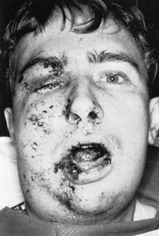
For soft-tissue injuries to the face, the goal is to retain function and have good cosmesis. It is important that repair of facial wounds occur is a timely manner. Because of the highly vascular nature of the face, the length of time for wound closure can be extended to 20 hours from the time of injury, although it is preferable to delay no longer than 8 to 12 hours. In a healthy patient the face is considered to be at low risk for infection. Deeper lacerations and lacerations associated with fractures can be conservatively debrided, irrigated, and closed before reduction. With tissue that is considered viable, excessive debridement should be avoided. Repair of facial lacerations in uncooperative patients is extremely difficult and may injure other important structures. Delaying repair until the patient is more cooperative usually results in better outcome. Figure 27-1 shows contusions, abrasions, and lacerations of the face.
 |
| FIGURE 27-1 Facial injuries. (From Danis DM, Blansfield JS, Gervasini AA: Manual of clinical trauma care: the first hour, ed 4, St. Louis, 2007, Mosby.) |
Lacerations caused by animal or human bites are highly contaminated because of the bacteria and debris found in the mouth. All bites should be meticulously cleaned with soap and water and irrigated with warmed saline (preferred) because the warm temperature is more appealing to the patient. Detergent, hydrogen peroxide, and concentrated povidone-iodine solutions should be avoided because they are considered toxic to the tissues. 8 There are many factors to consider when deciding whether to close a facial laceration caused by a bite. Human and animal bite wounds on the face can be disfiguring, so suturing is more commonly done; however, cat bites, which are most often puncture wounds, are left open. Extensive or gaping wounds on the face present cosmetic problems, so consultation with a plastic surgeon is recommended. Most experts recommend closing the wound after meticulous irrigation and debridement. Both human and animal bites should be inspected for tooth fragments. Extensive animal bites, usually caused by large dogs, frequently require surgical exploration and repair. A helpful mnemonic for dealing with animal bites is RATS (rabies, antibiotics, tetanus, and soap). All patients should be covered for tetanus and rabies prophylaxis as indicated. Patients with animal and human bites should receive prophylactic antibiotics that cover streptococcus, staphylococcus, Eikenella corrodens, and Pasteurella multocida. 8
Stay updated, free articles. Join our Telegram channel

Full access? Get Clinical Tree


Get Clinical Tree app for offline access
