Section Twenty-Three Dental and Throat Procedures
PROCEDURE 173 Indirect Laryngoscopy
CONTRAINDICATIONS AND CAUTIONS
1. If possible, epiglottitis should be ruled out before attempting indirect laryngoscopy.
2. Indirect laryngoscopy has been almost replaced by the use of flexible fiberoptic laryngoscopes. Skillful use of a head mirror and light provides a wide field of view, light which is coaxial with vision, and more work space. Flexible or rigid endoscopy gives more detailed inspection, can be better directed, may allow railroading of a tube, possible video recording, and has less gagging with nasal passage but increases technical complexity and disinfection requirements and is difficult with increased secretions.
EQUIPMENT
Laryngeal mirror (size 3 to 6)
Light source, such as a headlamp or head mirror
A method to prevent fogging (i.e., warming the mirror with warm water, an alcohol lamp, or an electric light bulb)
Antifogging solution (commercially available solution or soapy water can be used)
Topical anesthetic (e.g., lidocaine, Cetacaine, or aerosolized tetracaine)
PATIENT PREPARATION
1. Position the patient sitting upright with feet on the floor. Have the patient lean forward with the head in the sniffing position.
2. Place an emesis basin in the patient’s hands.
3. *Anesthetize the pharynx as indicated by patient response and cooperation (see Procedure 135 for information on topical anesthetics).
PROCEDURAL STEPS
1. Have the patient protrude the tongue from the mouth as far as possible.
2. *Lay gauze over the tongue and then wrap it under the tongue.
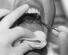
3. *Grip the gauze-wrapped tongue between the thumb and the index finger of the nondominant hand and brace the middle finger against the teeth; then elevate the upper lip (Figure 173-1).
4. *Warm the mirror and test the temperature on your hand.
5. *Slide the mirror base down carefully, keeping it parallel to the tongue and without touching any tissue.
6. *Place the mirror with the back side against the uvula.
7. *Elevate the uvula and soft palate using one motion.
8. *Avoid touching the posterior tongue; this stimulation may result in gagging.
9. Instruct the patient to concentrate on breathing normally through the mouth with eyes open and focused on a distant fixed object.
10. *Direct the light onto the mirror.
11. *Examine the structures; look for pathology or a foreign body.
12. *Ask the patient to say “E.” Observe the vocal cords as the epiglottis is displaced.
PROCEDURE 174 Esophageal Foreign Body Removal
The esophagus has three areas of narrowing: upper esophageal sphincter, which consists of the cricopharyngeus muscle; crossover of the aorta; and lower esophageal sphincter. These areas are where most esophageal foreign bodies become entrapped (Thomas & Brown, 2006). Structural abnormalities, including strictures, diverticula, and malignancies, increase the risk of foreign body entrapment, as do motor disturbances, such as scleroderma and achalasia. The oropharynx is well innervated, and patients can typically localize oropharyngeal foreign bodies; however, foreign bodies in the lower two thirds of the esophagus are poorly localized.
ESOPHAGOSCOPY
Indications
1. This method provides direct visualization of the foreign body and the ability to evaluate the esophagus for pathology and allows control of the object during removal. Esophagoscopy is the preferred method of removal for sharp objects (e.g., bones, safety pins, and razor blades) and button batteries that may rapidly cause esophageal injury.
2. Esophagoscopy may also be used to rule out predisposing pathology or resultant complications (Munter & Heffner, 2004). An emergency physician does not usually perform esophagoscopy; a specialist is consulted.
Contraindications and Cautions
Sharp, pointed objects (e.g., toothpicks, bones) may require removal in the operating room.
Patient Preparation
1. Establish venous access for medication administration (see Procedure 60).
2. If the object poses a high risk for esophageal perforation, prophylactic antibiotics should be administered intravenously before the endoscopy (Munter & Heffner, 2004).
3. Consider endotracheal intubation if the airway is at risk for compromise (see Procedures 8 through 11).
4. Place the patient in a Trendelenburg, lateral decubitus position.
5. Assess vital signs and oxygen saturation, and continue to monitor them frequently throughout the procedure (see Procedures 21 and 55).
6. *Administer a topical anesthetic as prescribed to control gagging.
7. Administer sedatives and analgesics as prescribed.
8. Have suction assembled and turned on at the patient’s head.
Procedural Steps
1. *Intubate the esophagus with the endoscope.
2. *Visualize the foreign body.
3. *Push the foreign body into the stomach; grasp it and remove it through the scope; or grasp it and remove it with the scope as a unit.
4. *Evaluate the esophagus for preexisting pathology or trauma induced by the foreign body.
Stay updated, free articles. Join our Telegram channel

Full access? Get Clinical Tree


