CHAPTER 22. Spinal Trauma
Jennifer Wilbeck
Trauma to the spinal column and the spinal cord can result in devastating and life-threatening injuries. Each year approximately 11,000 acute spinal cord injuries occur in the United States. The vast majority of those injured are males under age 38 years. Males have remained the gender most commonly injured for decades; however, the average age at injury has steadily increased over the past three decades as the same trend has been observed in the general United States population. The estimated cost for care ranges from $218,500 to more than $741,400 during the first year after the patient’s injury. Lifetime costs vary based on age of the patient and site of the injury. High cord injuries in young patients cost just under $3,000,000 per year. Although prehospital care, medical, surgical, and technologic advances have contributed to an increased life span for those who have suffered a spinal cord injury, the life expectancy of these patients remains lower than for patients with no spinal injury. 21
Spinal trauma can occur as a result of both blunt and penetrating trauma. The most common mechanism of injury is a motor vehicle crash, accounting for nearly half of acute spinal cord injuries. 21 Motor vehicle crashes cause spinal cord injuries through rollovers, occupant ejection, and collisions with pedestrians. Spinal trauma also commonly results from falls, acts of violence (including penetrating wounds from guns or knives), and sports injuries. In older adults, falls are the leading cause of trauma. 2 Although the number of spinal injuries due to falls is continually rising, the number of injuries resulting from violence has trended down since the mid-1990s. 16 Increased participation in extreme sports and outdoor activities such as in-line skating, snowboarding, and bicycling have also contributed additional sources for spinal cord injuries.
In the past, many people with spinal cord injuries died from respiratory complications such as aspiration and pneumonia. 34 Establishment of spinal cord injury care systems has decreased complications from spinal cord injuries and improved survival of those injured. Spinal cord patients need to be transferred to the appropriate definitive care facility as soon as possible to decrease complications and costs that may occur related to the injury.
Care of the patient with spinal trauma begins in the prehospital environment with rapid identification of actual injury or potential for injury based on the mechanism of injury, immediately followed by appropriate spinal protection interventions. Recent studies indicate that patients with head injuries are at higher risk for also having cervical spine injuries, particularly if the patient is unconscious or has a focal neurologic deficit. 11. and 14. On arrival in the emergency department (ED), the patient should be fully evaluated to rule out concomitant life-threatening injuries such as tension pneumothorax or intraabdominal bleeding while spine protection is continued.
Emergency care of the patient with spine trauma requires an organized, multidisciplinary approach. Patient survival and quality of life after the acute injury depend on the emergency care a patient receives. This chapter discusses anatomy and physiology of spine trauma, mechanisms of injury, patient assessment and initial interventions, specific injuries, and current research related to management of acute spine injury.
ANATOMY AND PHYSIOLOGY
Vertebral Column
The vertebral column serves as bony support for the head and trunk and provides protection for the spinal cord. A total of 7 cervical vertebrae, 12 thoracic vertebrae, 5 lumbar vertebrae, 1 sacral vertebra (composed of 5 fused vertebrae), and 1 coccygeal vertebra (composed of 4 fused vertebrae) constitute the vertebral column. Each vertebra is composed of a body, a vertebral arch, and a vertebral foramen. The arch of the vertebra is composed of two pedicles, two laminae, four articular processes (facets), two transverse processes, and the spinous process, which can be felt when palpating the posterior spine. 27. and 33.
The cervical vertebrae are the most frequently injured vertebrae because they are the most mobile part of the spine and because they are so small and delicate. 27 The rib cage provides stability to the vertebrae from T-1 to T-10 and keeps this portion of the spine relatively immobile. Because the thoracic vertebrae are so strong, fractures and/or dislocations at this level should increase suspicion for spinal cord injury. 19 The lumbar vertebrae are the largest and strongest in the vertebral column. 27. and 33.
Ligaments attach to the transverse and spinous processes to connect the vertebral bodies and provide support and stability to the vertebral column. They also limit the spinal column from excessive flexion and extension. Between the vertebral bodies are discs that act as shock absorbers and articulating surfaces for the adjacent vertebral bodies. 33. and 35.
Spinal Column
The spinal cord extends from the brain, through the foramen magnum, and down the vertebral column to the level of L2. This mass of nerve tissue regulates body movement and function through transmission of nerve impulses. The diameter of the spinal cord is largest in the cervical and lumbar regions and tapers in the lower thoracic area. In adults it terminates in a cone-shaped structure, known as the conus medullaris, at the L1 or L2 level. Spinal nerve roots that exit below the conus medullaris are referred to as the cauda equina. 5.27. and 31.
The primary function of the spinal cord is to regulate bodily function and movement by transmitting nerve impulses between the brain and the body. Cross-sectional views of the spinal cord reveal a butterfly-shaped core composed of gray matter, surrounded on the outer edges by white matter. The gray matter contains nerve cell bodies and is divided into three distinct regions, each with specific characteristics: the posterior (dorsal), intermediolateral (lateral), and anterior (ventral) horns. The posterior, or dorsal, horn contains sensory interneurons and axons whose cell bodies are located in the dorsal root ganglion. The intermediolateral, or lateral, horn contains cell bodies with autonomic nervous system function. The anterior, or ventral, horn contains somatic motor neurons that leave the spinal cord via the spinal nerves. 27. and 31.
The white matter of the spinal cord consists of multiple ascending and descending pathways (referred to collectively as “spinal tracts”), which are individually named based on their origins and terminations. These tracts run parallel to the spinal cord’s vertical axis and transmit action potentials to and from the brain to other parts of the spinal cord. Table 22-1 lists some of these specific tracts and describes their functions. 27. and 31.
| Spinal Tract | Function |
|---|---|
| Dorsal column (ascending) | Proprioception, pressure, and vibration |
| Lateral spinothalamic tract (ascending) | Pain and temperature |
| Anterior spinothalamic tract (ascending) | Light touch, pressure, and itch sensation |
| Spinocerebellar tract (ascending) | Proprioception to the cerebellum |
| Pyramidal tracts (descending) | Voluntary control of skeletal muscle |
| Extrapyramidal tracts (descending) | Automatic control of skeletal muscle |
Spinal Nerves
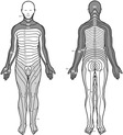
The spinal cord has 31 pairs of spinal nerves that exit the spinal cord bilaterally and provide pathways for involuntary responses to specific stimuli. There are 8 cervical nerves, 12 thoracic nerves, 5 lumbar nerves, 5 sacral nerves, and 1 coccygeal nerve. The spinal nerves innervate voluntary striated muscle and are responsible for the majority of the communication between the spinal cord and the rest of the body. Each of these nerves has a posterior root that transmits sensory impulses from the periphery into the spinal cord and an anterior root that transmits motor impulses from the spinal cord out to the periphery. Table 22-2 lists the spinal nerve muscle innervations and their corresponding expected patient response. The dorsal root of these nerves innervates a distinct region of the body surface known as a dermatome. 27. and 33. Assessment of the 28 dermatomes provides information about function to sensory areas of the spinal cord. Figure 22-1 illustrates the sensory dermatomes.
| Nerve Level | Muscles Innervated | Patient Response |
|---|---|---|
| C4 | Diaphragm | Ventilation |
| C5 | Deltoid | Shrug shoulders |
| Biceps | Flex elbows | |
| Brachioradialis | ||
| C6 | Wrist extensor | Extend wrist |
| Extensor carpi radialis longus | ||
| C7 | Triceps | Extend elbow |
| Extensor digitorum communis | Extend fingers | |
| Flexor carpi radialis | ||
| C8 | Flexor digitorum profundus | Flex fingers |
| T1 | Hand intrinsic muscles | Spread fingers |
| T2 to T12 | Intercostals | Vital capacity |
| L1 | Abdominal | Abdominal reflexes |
| L2 | Iliopsoas | Hip flexion |
| L3 | Quadriceps | Knee extension |
| L4 | Tibialis anterior | Ankle dorsiflexion |
| L5 | Extension hallucis longus | Ankle eversion |
| S1 | Gastrocnemius | Ankle plantar flexion |
| Big toe extension | ||
| S2 to S5 | Perineal sphincter | Sphincter control |
 |
| FIGURE 22-1 Sensory dermatomes. (From Marx JA, Hockberger RS, Walls RM, et al: Rosen’s emergency medicine: concepts and clinical practice, ed 6, St. Louis, 2006, Mosby.) |
Vascular Supply
The vascular supply for the spinal cord comes from branches of the vertebral arteries and the aorta. The anterior and posterior spinal arteries branch off the vertebral artery at the cranial base and descend parallel to the spinal cord. 31 Because spinal cord arteries cannot develop collateral blood supply, injuries to these arteries can be devastating.
PATIENT ASSESSMENT
The emergency nurse should perform primary and secondary assessments of injured patients while simultaneously performing interventions to stabilize the airway, breathing, and circulation. All patients with multisystem injuries or significant mechanisms of injury must be suspected of having a spinal injury and should be completely immobilized. Spinal protection involves manual immobilization of the patient’s head until a rigid cervical collar, lateral head support such as head blocks or rolled sheets, and backboard have been applied. The entire spine should be immobilized using a backboard with straps across the chest, abdomen, and knees. 11.14. and 18.
Once the initial assessment and resuscitation have been completed, the patient should be removed from the backboard as soon as safely possible. Patients who have sustained spinal trauma, particularly those with sensory losses, are at great risk for developing skin breakdown, putting the patient at increased risk for additional injury and infection. 14. and 20.
After the patient’s critical needs have been met, the emergency nurse may then perform a more focused assessment related to spinal injury. The initial assessment includes obtaining mechanism of injury and other history, as well as performing a focused examination of the spine and spinal cord.
Mechanisms of Injury
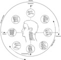
Mechanisms of spine injury include blunt and penetrating forces. The vertebrae, spinal cord, and nerve roots may be injured as a result of fractures, dislocations, or subluxation. The cord may also be injured through direct penetration by a bullet, knife, or other sharp object, or by adjacent injuries resulting in localized edema that compresses the spinal cord. Six basic types of movement can injure the spinal cord. These are illustrated in Figure 22-2 and summarized in Table 22-3.
 |
| FIGURE 22-2 Mechanisms of injury to the spine. The mechanism of cervical injury (flexion versus extension) determines the type of cervical spine fracture or dislocation. (From Moore EE, editor: Early care of the injured patient, ed 3, Philadelphia, 1990, Decker.) |
| Category | Mechanism of Injury |
|---|---|
| Hyperextension | The head is forced back, and the vertebrae of the cervical region are placed in an overextended position. |
| Hyperflexion | The head is forced forward, and the cervical vertebrae are placed in an overflexion position. |
| Axial loading | A severe blow to the top of the head causes a blunt downward force on the vertebrae and the spinal column. |
| Compression | Forces from above and below compress the vertebrae. |
| Lateral bend | The head and neck are bent to one side, beyond the normal range of motion. |
| Overrotation and distraction | The head turns to one side, and the cervical vertebrae are forced beyond normal limits. |
The following mechanisms have been identified as being commonly associated with cervical spine injuries: fall from a height greater than or equal to 3 feet or five stairs; axial loading; motor vehicle collision at a high rate of speed with rollover or ejection; collision involving a motorized recreational vehicle; and bicycle collisions. 30 In addition, spinal injuries should be suspected if a fall results in fractures of the heels7 or if an unrestrained (no seat belt) patient presents with facial injuries.
History
In addition to the mechanism of injury, the following pieces of history may indicate a potential spinal cord injury: history of significant trauma and altered mental status from intoxication; history of seizure activity since the incident; any complaint of neck pain or altered sensation in the upper extremities; complaint of neck tenderness; history of loss of consciousness; injuries to the head or face. When obtaining the history, the emergency nurse should ask the patient about neck pain and changes in sensation or movement since the time of injury. Any history of incontinence before arrival in the ED is important to identify because it may signal a spinal cord injury. 11.18. and 30.
If the patient is unconscious, prehospital history may be the only source of information available concerning the patient’s condition and behaviors immediately after the incident; thus the emergency nurse should obtain as much information from prehospital personnel as possible. Patients who are unconscious or have any altered mental status and those with distracting injuries are at a higher risk for missed cervical spine injury because their injuries are more easily overlooked and they are unable to report any indicative symptoms. 14. and 30.
Inspection
The emergency nurse should observe the patient for obvious signs of spinal injury, including deformity of the vertebral column, cervical edema, and ballistic wounds in the neck, chest, or abdomen. Abrasions or contusions at the level of the lap or shoulder belt in restrained patients can indicate spinal injuries at those levels. 14
The patient’s ventilatory pattern and effort may also indicate a cord injury. Injuries to the spinal cord between C3 and C5 can result in progressive respiratory insufficiency secondary to significant respiratory muscle dysfunction when diaphragmatic innervation by the phrenic nerve is altered. Injuries to the cord below C5 lead to decreased intercostal and abdominal muscle function, in turn altering the normal mechanics of ventilation and increasing the work of breathing. 5.18. and 34.
The patient’s ability to move and perceive pain during procedures such as arterial punctures or intravenous catheter insertion is another important observation. The patient should be able to wiggle his or her toes and fingers and to lift arms and legs. The inability to do so indicates some type of spinal injury. Continued penile erection (priapism) can occur with loss of sympathetic nervous system control and may indicate a cervical spine injury. 19
Leakage of cerebral spinal fluid (CSF) from the nose or ears may also be noted. Confirm the presence of CSF using the halo test (as discussed in Chapter 21). Due to the high incidence of simultaneous closed head injuries and cervical spine injuries, patients with CSF leaks raise even higher suspicion for serious spinal trauma.
Palpation
Injuries above the T4 level usually disrupt the sympathetic nervous system, causing vasodilation below the level of the injury. If the patient is diaphoretic, sweat is present above rather than below the level of the injury. In addition, a patient with a spinal cord injury becomes poikilothermic, assuming the temperature of his or her surroundings, because of loss of sympathetic tone. This can leave the patient at great risk for becoming hypothermic. 5
Pulse rate and quality should be palpated. In neurogenic shock the pulse is slow and strong, whereas in hypovolemic shock it is rapid and weak. 5
Strength and symmetry of movement in all four extremities should be evaluated (see Table 22-2). A quick motor evaluation should include flexion and extension of the arms, flexion and extension of the legs, flexion of the foot, extension of the toes, and sphincter tone.
Sensory status may be assessed by evaluation of dermatomes. The patient should be able to distinguish between sharp and dull sensations using a safety pin or cotton swab. Testing should begin at the level of no reported sensation and proceed upward to identify the level at which feeling returns.
The presence of sacral or perineal sensations should also be assessed. 14 If sacral sensations are present in patients with other focal deficits (termed “sacral sparing”), an incomplete spinal cord injury should be suspected.
Finally, the patient’s entire spinal column should be gently palpated for pain, tenderness, crepitus, and step-off deformity. Palpation requires that the patient be logrolled by at least three team members to maintain spinal alignment. 14 If the cervical collar is removed for this procedure, manual stabilization must be maintained.
Reflex Testing
The emergency nurse may perform an assessment of the patient’s reflexes. Reflexes are summarized in Table 22-4.
| Reflex | Spinal Cord Level |
|---|---|
| Biceps | C5-C6 |
| Brachioradialis | C5-C6 |
| Triceps | C6-C7 |
| Superficial abdominal (above umbilicus) | T8-T10 |
| Superficial abdominal (below umbilicus) | T10-T12 |
| Knee jerk | L2-L4 |
| Ankle | S1 |
| Anal wink | S2-S4 |
| Plantar response | L5-S1 |
Stay updated, free articles. Join our Telegram channel

Full access? Get Clinical Tree


Get Clinical Tree app for offline access
