Section Twenty-One Otic Procedures
PROCEDURE 162 Instillation of Ear Medications
PROCEDURE 163 Ear Wick Insertion
PROCEDURE 165 Otic Foreign Body Removal
PROCEDURE 162 Instillation of Ear Medications
CONTRAINDICATIONS AND CAUTIONS
1. Caution should be exercised to identify perforation of the tympanic membrane before instilling medications into the external auditory canal, because inadvertent introduction of foreign material into the middle ear may result in an infection.
2. Medications in suspension (oil based) do not absorb into ear wicks. Use ear wicks with medication in solution form only.
PROCEDURAL STEPS
1. Place the patient on his or her side with the affected ear up.
2. Straighten and examine the external auditory canal. For an infant, gently pull the pinna down and back. For a child older than age 3 or an adult, pull the pinna up and backward (see Figure 164-1).
3. Clear any debris from the ear canal (see Procedures 164 through 166).
4. Warm the ear medication container by holding it in your hand or placing it in warm water for a short time.
5. Draw up only the amount of medication needed into the ear dropper. Do not return excess medication to the bottle.
6. Instill the correct number of drops along the side of the ear canal. To prevent contamination, do not allow the tip of the ear dropper to touch any part of the ear. Hold it about ½ inch above the ear canal.
7. Press firmly but gently on the tragus of the ear a few times. This helps the medication reach all portions of the canal.
8. Have the patient remain in the same position (with the affected ear up) for a few minutes.
9. Place a small piece of a cotton ball at the external auditory meatus for 15 minutes to help retain the medication when the patient is up. Do not press the cotton into the canal.
10. If an ear wick is to be used, see Procedure 163. Saturate the wick with a topical otic solution.
COMPLICATIONS
1. If cotton balls are used instead of an ear wick, disintegration may occur, and removal may be difficult. To avoid retention of pieces of cotton balls, place them only at the meatus.
2. Without an ear wick or a cotton ball, solutions or suspensions may extravasate from the ear canal.
3. Otic solutions frequently cause a burning or stinging sensation; suspensions are less painful.
PATIENT TEACHING
1. Report immediately ear pain, an edematous canal, tender ear cartilage, purulent drainage, or hearing loss.
2. If present, leave the ear wick in place for 48 to 72 hours. Repeat instillation of medication onto the ear wick three or four times daily or as directed.
3. Continuation of medication may be necessary after ear wick removal (approximately 10 days for antibiotics).
4. If an ear wick is not used, you may place a cotton ball at the external auditory meatus to prevent extravasation of the medication.
PROCEDURE 163 Ear Wick Insertion
PROCEDURAL STEPS
1. Using an otoscope, check the external ear for redness or drainage.
2. Examine the internal ear with an otoscope.
3. Suction debris from the ear with the small metal suction tip, or remove debris gently with a cerumen spoon.
4. Irrigation may be necessary (after visualization of ear drum) before placement of ear wick to allow effective dispersal of medications to inflamed tissue. See Procedure 164.
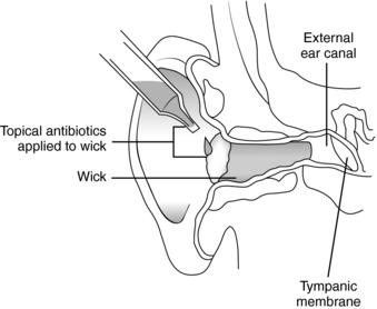
5. Use a small alligator forceps to twist the ear wick in place in the canal. Follow the accompanying directions to insert a commercially prepared wick, or make a wick by wrapping cotton or selvedged gauze tightly around the tip of an alligator forceps, grasping the cotton or gauze with the forceps, and placing it in the ear canal. Antibiotic or steroid cream can be applied to gauze before wrapping.
6. Place the prescribed ear drops in the ear canal, taking care not to touch the dropper to the ear canal (Figure 163-1). Place drops along the side of the canal so that air is displaced as they flow. Cold drops may cause vertigo or nausea; this can be avoided by warming the medication slightly in your hands or by immersing the bottle in a cup of warm water for several minutes.
7. Place 1 or 2 additional drops on the ear wick to saturate it.
PATIENT TEACHING
1. Most ear wicks will spontaneously extrude in 12 to 36 hours (Block, 2005). If necessary, return in 24 to 48 hours to have ear wick removed or follow up with your physician to have it removed. Make sure the wick is moist before removing it.
2. Instill ear drops as instructed. Cold ear drops may cause dizziness or nausea. Warm the bottle between your hands or in a cup of warm water before instilling the drops.
3. Symptoms should subside in 1 or 2 days. If no improvement occurs or if symptoms worsen, contact your physician.
4. To prevent the ear wick from slipping, do not touch the ear, and do not place cotton in the ear canal over the wick, because the cotton will absorb all the drops.
5. Take oral pain medications as prescribed and apply a warm moist compress to ear for pain.
6. To prevent future infections, do not put foreign objects in the ear. Ear rinses used after swimming may be helpful. Avoid strong jets from showerheads going directly into the ears.
PROCEDURE 164 Ear Irrigation
CONTRAINDICATIONS AND CAUTIONS
1. Irrigation is contraindicated if the tympanic membrane is perforated or potentially not intact, (secondary to injury, myringotomy tubes, or surgery) or in the presence of severe external otitis (Riviello, 2004).
2. Avoid extreme temperatures, which may cause pain, dizziness, nausea, and vomiting (Riviello, 2004).
3. If there is water-absorbent material in the ear, such as vegetable matter (e.g., beans), do not irrigate, because the material may swell and make removal more difficult (see Procedure 165) (Riviello, 2004).
4. Be careful not to abrade the ear canal with the irrigating device.
5. Discontinue the procedure and notify the physician if the patient experiences pain, vertigo, or nystagmus (Parshall, 2005).
6. Use caution with elderly and immunocompromised patients because irrigation may lead to malignant otitis externa (Riviello, 2005).
7. Do not attempt on an uncooperative patient or child who cannot be still or adequately restrained to perform the procedure safely (Forzley, 2003).
8. Referral to a specialist should be considered if the affected ear is the only hearing ear (Forzley, 2003).
EQUIPMENT
Otologic ear syringe (metal ear syringe)
60-ml syringe with an 18- or 20-G intravenous catheter sheath attached, or a butterfly, with the needle and wings removed, cut to leave approximately 1 in of tubing near the hub
Dental irrigating device (such as a Water-Pik) on low setting (A proper ear irrigating syringe is preferred to extemporized devices or a Water-Pik because it supplies a large volume at low pressure, rather than the high pressures of other devices.)
Irrigant—warm water, warm normal saline, or half-strength hydrogen peroxide
Cotton balls or gauze dressings
Stay updated, free articles. Join our Telegram channel

Full access? Get Clinical Tree


