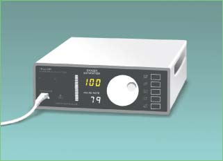
Pulse Oximetry
Performed intermittently or continuously, pulse oximetry is a relatively simple procedure used to monitor arterial oxygen saturation noninvasively. Pulse oximeters usually denote arterial oxygen saturation values with the symbol SpO2, whereas invasively measured arterial oxygen saturation values are denoted by the symbol SaO2.
In this procedure, two diodes send red and infrared light through a pulsating arterial vascular bed, like the one in the fingertip. A photodetector slipped over the finger measures the transmitted light as it passes through the vascular bed, detects the relative amount of color absorbed by arterial blood, and calculates the arterial oxygen saturation without interference from surrounding venous blood, skin, connective tissue, or bone. Using the ear probe, oximetry works by monitoring the transmission of light waves through the vascular bed of a patient’s earlobe. Results will be inaccurate if the patient’s earlobe is poorly perfused, as from a low cardiac output. (See How oximetry works, page 618.)
Equipment
Oximeter ▪ finger or ear probe ▪ alcohol pads ▪ nail polish remover, if necessary.
Implementation
Confirm the patient’s identity using at least two patient identifiers according to your facility’s policy.4
Explain the procedure to the patient. Answer all questions to decrease anxiety and increase cooperation.
For Pulse Oximetry Using A Finger Probe
Select a finger for the test. Although the index finger is commonly used, a smaller finger may be selected if the patient’s fingers are too large for the equipment. Make sure the patient isn’t wearing false fingernails, and remove any nail polish from the test finger. Place the transducer (photodetector) probe over the patient’s finger so that light beams and sensors oppose each other (as shown below). If the patient has long fingernails, position the probe perpendicular to the finger, if possible, or clip the fingernail. Always position the patient’s hand at heart level to eliminate venous pulsations and to promote accurate readings.
 |
How Oximetry Works
The pulse oximeter (shown below) allows noninvasive monitoring of the percentage of hemoglobin saturated by oxygen, or SpO2 levels, by measuring the absorption (amplitude) of light waves as they pass through areas of the body that are highly perfused by arterial blood. Oximetry also monitors pulse rate and amplitude.
 |
Light-emitting diodes in a transducer (photodetector) attached to the patient’s body (shown below on the index finger) send red and infrared light beams through tissue. The photodetector records the relative amount of each color absorbed by arterial blood and transmits the data to a monitor, which displays the information with each heartbeat. If the SpO2 level or pulse rate varies from preset limits, the monitor triggers visual and audible alarms.
Stay updated, free articles. Join our Telegram channel

Full access? Get Clinical Tree


Get Clinical Tree app for offline access
