Pulse Assessment
Blood pumped into an already-full aorta during ventricular contraction creates a fluid wave that travels from the heart to the peripheral arteries. This recurring wave—called a pulse—can be palpated at locations on the body where an artery crosses over bone on firm tissue. In adults and children older than age 3 and in adults with a suspected cardiac disorder that affects the patient’s heart rate and rhythm, the radial artery in the wrist is the most common palpation site. (See Pulse points.) In infants and children
younger than age 3, a stethoscope is used to listen to the heart itself rather than palpating a pulse. Because auscultation is done at the heart’s apex, this is called the apical pulse.
younger than age 3, a stethoscope is used to listen to the heart itself rather than palpating a pulse. Because auscultation is done at the heart’s apex, this is called the apical pulse.
An apical-radial pulse is taken by simultaneously counting apical and radial beats—the first by auscultation at the apex of the heart, the second by palpation at the radial artery. Some heartbeats detected at the apex can’t be detected at peripheral sites. When this occurs, the apical pulse rate is higher than the radial; the difference is the pulse deficit.
Pulse taking involves determining the rate (number of beats per minute), rhythm (pattern or regularity of the beats), and volume (amount of blood pumped with each beat). If the pulse is faint or weak, use a Doppler ultrasound blood flow detector if available.
Equipment
Watch with second hand ▪ stethoscope (for auscultating apical pulse) ▪ alcohol pad.
Preparation of Equipment
If you aren’t using your own stethoscope, disinfect the earpieces with an alcohol pad before and after use to prevent cross-contamination.1
Implementation
Confirm the patient’s identity using at least two patient identifiers according to your facility’s policy.5
Tell the patient that you intend to take his pulse.
Make sure the patient is comfortable and relaxed because an awkward, uncomfortable position may affect the heart rate.
Taking A Radial Pulse
Place the patient in a sitting or supine position, with his arm at his side or across his chest.
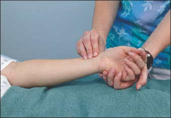
Gently press your index, middle, and ring fingers on the radial artery, inside the patient’s wrist (as shown below). You should feel a pulse with only moderate pressure; excessive pressure may obstruct blood flow distal to the pulse site. Don’t use your thumb to take the patient’s pulse because your thumb’s own strong pulse may be confused with the patient’s pulse.

After locating the pulse, count the beats for 60 seconds, or count for 30 seconds and multiply by 2. Counting for a full minute provides a more accurate picture of irregularities. While counting the rate, assess pulse rhythm and volume by noting the pattern and strength of the beats. If you detect an irregularity, repeat the count, and note whether it occurs in a pattern or randomly. If you’re still in doubt, take an apical pulse. (See Identifying pulse patterns, page 616.)
Stay updated, free articles. Join our Telegram channel

Full access? Get Clinical Tree


Get Clinical Tree app for offline access
