Peripherally Inserted Central Catheter Use
A peripherally inserted central catheter (PICC) is a catheter that’s inserted percutaneously into a peripheral vein. The catheter tip resides in the lower one-third of the superior vena cava, at the junction of the superior vena cava and right atrium. PICC placement is indicated for intermediate to long-term IV access for the administration of antibiotics, pain medications, chemotherapy or other vesicants, parenteral nutrition or other hyperosmolar solutions, or blood products. The early use of PICCS may also spare peripheral veins, limit the pain of repeated needle sticks, and help avoid complications that may occur with a central venous (CV) access device. PICCs are also used for patients receiving home care.
PICCs are made of silicone or polyurethane and vary in diameter and length. They’re available in single, double, and triple-lumen versions, with and without valves. The type and size of PICC used depends on the patient’s size and anatomic measurements as well as the therapy required; the patient receiving PICC therapy must also have a peripheral vein large enough to accept an introducer needle and catheter.
Most PICCs are manufactured with smooth, rounded tips to reduce trauma to the vein wall during insertion. Selecting a catheter length that’s as close to the desired patient length measurement as possible avoids the need to trim the PICC (Groshong PICCs can’t be trimmed). The catheter length should be subtracted from the total length of the PICC, with any extra length left outside the insertion site, safely secured.
Most PICCs have a preloaded stylet wire inside the catheter to add stiffness to the soft catheter, easing its advancement through the vein. The stylet terminates 1 to 2 cm away from the catheter tip. If the catheter length does require trimming, the stylet must be withdrawn and repositioned 1 to 2 cm from the cut end. The stylet should never be cut; it’s removed after insertion.
PICC devices are easier to insert than other CV devices. A single catheter may be used for the entire course of therapy, resulting in greater convenience and reduced cost. A nurse trained in PICC insertion may perform the procedure if the state nurse practice act permits it, although the nurse may have to demonstrate competence every year.
Possible veins for PICC insertion include the basilic, median cubital, cephalic, and brachial. When selecting a site, avoid areas that are painful on palpation; veins compromised by bruising, infiltration, phlebitis, sclerosing, or cording; and, in patients with stage 4 or 5 chronic kidney disease, forearm and upper arm veins that could be used for vascular access. In a patient who has had breast surgery with axillary node dissection, don’t use arm veins on the affected side; also avoid veins on the affected side in stroke patients, those undergoing radiation therapy, and those with lymphedema.1 Use an ultrasound device to guide insertion to increase your rate of success and reduce the risk for insertion-related complications.2 (See Bedside ultrasound for PICC insertion.) When inserted, the catheter is advanced until it reaches the superior vena cava near the junction of the right atrium.2 To help prevent catheter-related bloodstream infections, use maximum sterile barriers during insertion.3
PICCs may be inserted with a through-the-introducer or modified Seldinger technique. The procedure below discusses the modified Seldinger technique.
PICC infusion and associated site care, including flushing of the catheter, may be performed by a nurse or appropriately trained parent or guardian. A PICC dressing should be changed at least every 7 days if a transparent semipermeable dressing is used.2,4 If the patient is diaphoretic or the site is bleeding or oozing, a gauze dressing should be used instead of a semipermeable dressing; gauze dressings should be changed every 2 days. Replace either dressing if it becomes damp, loosened, or visibly soiled.2,3
PICCs are often used for administration of drugs, such as opioids, analgesics, antibiotics, parenteral nutrition, and chemotherapy. To prevent catheter-related bloodstream infections, wipe the injection surface with alcohol using friction and let it air-dry each time you access it.3
PICCs are removed when therapy is complete, if the catheter becomes damaged or broken and can’t be repaired, if the patient has discomfort and pain, or, possibly, if the catheter becomes occluded. Measure the catheter after you remove it to ensure that the catheter has been removed intact.5
Equipment
For Insertion
PICC insertion kit ▪ PICC catheter ▪ PICC insertion checklist ▪ chlorhexidine swabs ▪ sterile and clean measuring tape ▪ vial of normal saline solution ▪ syringes and needles of appropriate size ▪ sterile gauze pads ▪ linen-saver pad ▪ sterile drapes ▪ disposable skin marker ▪ tourniquet ▪ sterile marker ▪ sterile labels ▪ gloves ▪ sterile gloves ▪ sterile gown ▪ mask ▪ protective eyewear ▪ head cover ▪ 1% lidocaine without epinephrine ▪ catheter securement device, sterile tape, or sterile surgical strips ▪ sterile transparent semipermeable dressing ▪ Optional: bedside ultrasound equipment, sterile and clean disposable ultrasound probe cover, locator system, echogenic needle, heparin (10 units/mL).
Commercially prepared PICC insertion kits contain most of the equipment required to insert a PICC.
Bedside Ultrasound for PICC Insertion
Although special training is required, the use of bedside ultrasound helps provide safer, more efficient insertion of a peripherally inserted central catheter (PICC). Since the introduction of ultrasound, placing PICCs using visualization and palpation alone is rare.
Advantages of using ultrasound include:
the ability to locate the exact position of veins that are neither visible nor palpable and detect possible anatomic variations or thrombosis in the vessel
a successful cannulation rate of more than 90% on the first attempt
the possibility of inserting a PICC in a location away from the antecubital fossa, which can limit or eliminate complications such as mechanical phlebitis
a reduction in the complications related to traumatic placement.
For Flushing
Gloves ▪ syringe prefilled with appropriate flush solution6 (heparinized saline or preservative-free normal saline solution as ordered) ▪ antiseptic pads (alcohol, tincture of iodine, or chlorhexidine-based).
For Dressing Change
Gloves ▪ sterile gloves ▪ masks ▪ sterile drape ▪ 2% chlorhexidine swabs ▪ transparent semipermeable dressing ▪ sterile disposable tape measure ▪ sterile tape, sterile surgical strips, or securement device ▪ label ▪ Optional: adhesive remover, chlorhexidine sponge disk, povidone-iodine swabs, 70% alcohol swabs, skin preparation adhesive, 2″ × 2″ gauze pads.
Many facilities stock a commercially prepared or facility-prepared sterile dressing change kit that contains the necessary supplies.
For Drug Administration
Patient’s medication record ▪ gloves ▪ medication to be administered in an IV container with administration set (for infusion) or in a syringe (for IV bolus) ▪ two 10-mL syringes prefilled with normal saline solution ▪ heparin flush ▪ alcohol wipes.
For Removal
Gloves ▪ linen-saver pad ▪ sterile occlusive dressing ▪ antiseptic ointment ▪ tape ▪ measuring tape ▪ warm, moist pack ▪ Optional: personal protective equipment.
Implementation
Verify the doctor’s order.
Confirm the patient’s identity using at least two patient identifiers according to your facility’s policy.10
Explain the procedure to the patient and answer all questions to decrease anxiety and increase cooperation.
Inserting a PICC
Make sure the doctor has obtained an informed consent and that it’s documented in the medical record.11,12
Conduct a preprocedure verification process to make sure that all relevant documentation, related information, and equipment are available and correctly identified to the patient’s identifiers.13
Check the patient’s allergy status to prevent anaphylaxis.
If you’re using ultrasound, place a disposable cover on the bedside ultrasound probe. Then place a disposable tourniquet on the patient’s selected upper arm and examine it for appropriate veins. Mark the expected insertion site with a disposable skin marker and remove the tourniquet.
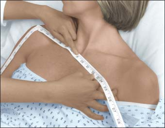
Using the clean measuring tape, determine catheter tip placement (the spot at which the catheter tip will rest after insertion).
For placement in the superior vena cava, measure the distance from the insertion site to the shoulder and from the shoulder to the sternal notch (as shown below).

Measure the midarm circumference of the selected extremity to provide a baseline measurement.
Have the patient lie in a supine position with her arm at a 90-degree angle to her body. Place a linen-saver pad under her arm.
Wash your hands for 60 seconds with antimicrobial soap and put on a surgical head cover, a gown, protective eyewear, and a mask.14,15
Prepare a sterile field using a sterile drape and set up the PICC supplies on the sterile field.
Prepare the lidocaine and normal saline flush syringes. Label all medications, medication containers, and other solutions on and off the sterile field using sterile marker and labels.16
If using ultrasound, ensure that a sterile probe cover is on the ultrasound probe to prevent cross-contamination between patients. If using a locator system, follow the manufacturer’s directions regarding its use.
Conduct a time-out immediately before starting the procedure to determine that the correct patient, site, positioning, and procedure are identified, and confirm, as applicable, that relevant information and necessary equipment are available.17
Prepare the insertion site by scrubbing with chlorhexidine swabs using a back-and-forth motion for about 30 seconds. Allow the area to dry. Don’t touch the intended insertion site.2
Place a full body drape over the patient from head to toe. Cover everything except the insertion site.
Place caps on the hub of each lumen of the catheter and prime each port. Always prepare the catheter according to the manufacturer’s recommendations.
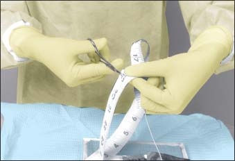
If it’s necessary to trim the PICC, pull the stylet wire back from the catheter tip. Using the sterile measuring tape, cut the distal end of the catheter to the appropriate premeasured length (as shown below).18 Follow the manufacturer’s recommendations and guidelines using the cutting equipment provided by the manufacturer. Then advance the stylet to 1 to 2 cm from the tip of the catheter and bend the stylet wire at the hub to prevent movement of the wire, which could result in trauma upon insertion. Place the catheter on the sterile field.

Remove and discard your gloves. Then apply the tourniquet again about 4″ (10 cm) above the antecubital fossa.
Put on a new pair of sterile gloves. Then place a sterile drape under the patient’s arm, and drape the arm with a large sterile drape. Drop a sterile 4″ × 4″ gauze pad over the tourniquet.
If using ultrasound, apply sterile ultrasound gel to the probe and use it to locate the appropriate vein.
Anesthetize the area with lidocaine to provide for patient comfort during insertion.
Perform a venipuncture using the appropriate needle. (An echogenic needle is used with ultrasound guidance.)
Advance a guide wire 2″ to 4″ (5 to 10 cm) through the needle and secure it to prevent embolization.
Remove the needle. If necessary, make a small skin nick at the insertion site to facilitate the advancement of the introducer/dilator.
Thread the introducer/dilator over the guide wire until you’re sure the tip is well within the vein. After successful vein entry, you should see a blood return.
Carefully remove the guide wire and advance the introducer the rest of the way into the vein.
Remove the dilator while holding the introducer still. To minimize blood loss, try applying finger pressure on the vein just beyond the distal end of the introducer sheath or place a finger over the opening of the introducer.
Using sterile forceps, insert the catheter through the introducer into the vein and advance it 2″ to 4″.
Remove the tourniquet using a sterile 4″ × 4″ gauze pad.
If using a locator system, activate it now and follow the manufacturer’s directions. Advance the catheter at a slow, steady pace until it’s in position at the premeasured length.
Grasp the tabs of the introducer sheath, and flex them toward its distal end to split the sheath. Peel the introducer while pulling away from the insertion site.
Check for a blood return and flush the catheter with normal saline solution. If you’re heparin locking the catheter, instill 5 mL of 10 units/mL heparin or follow your facility’s policy.19
Clean the site and secure the catheter with a catheter securement device, sterile tape, or sterile surgical strips.20
If necessary, apply a sterile 2″ × 2″ gauze pad directly over the site and a sterile transparent semipermeable dressing over the gauze pad.4 Leave this dressing in place for 24 hours.
Label the dressing with the date, time, and your initials.4
Obtain a chest X-ray to verify proper placement.2
Flushing a PICC
Thoroughly disinfect the needleless injection port with an antiseptic pad using friction and allow it to dry.3,22
Attach the syringe of preservative-free normal saline solution, making sure that the clamp on the PICC is closed to prevent accidental air embolism.

Stay updated, free articles. Join our Telegram channel

Full access? Get Clinical Tree


Get Clinical Tree app for offline access
