Pediatric Respiratory Disorders


Pediatric Respiratory Disorders


PEDIATRIC RESPIRATORY PROCEDURES


Oxygen Therapy

Children with respiratory problems may receive oxygen therapy via nasal cannula, mask, face tent, endotracheal tube, or tracheostomy device. An Isolette or oxygen hood may also be used for infants and young children. See Procedure Guidelines 44-1, pages 1480 to 1482.


Mechanical Ventilation

Infants and children requiring mechanical ventilation need specialized care. These patients are typically treated in facilities that focus on providing a safe environment for technology-dependent children, with care provided by highly skilled nurses, respiratory therapists, and pediatricians. Specific nursing procedures and interventions and management of technology-dependent infants and children are beyond the scope of this text. General considerations are as follows.


Maintenance of a Patent Airway


  • Artificial airway options include nasotracheal, orotracheal, and tracheostomy. Endotracheal (ET) and tracheostomy tubes are available in several sizes, cuffed and uncuffed, for the pediatric population.


    • Several methods are available to determine the appropriate size, such as: ET tube size = age in years + 16 divided by 4 (consult facility policy).


  • The patient should be closely monitored for hypoxia and bradycardia during intubation.


    • Only experienced and highly trained and skilled practitioners should perform intubation.


    • Resuscitation and reintubation equipment, access to oxygen and suction equipment must always be readily available.


    • An appropriate-size self-inflating bag with reservoir for oxygenation and mask should remain with the patient.


  • Action should be taken to prevent dislodgement of the artificial airway (especially during movement of the child) and obstruction of the airway.


    • Pediatric ET tubes have small diameters and are easily obstructed by thick secretions.


    • Adequate humidification will loosen secretions and suctioning will prevent airway obstruction.


    • Frequent vital signs and respiratory assessments are necessary. Cardiorespiratory monitors, pulse oximeter, endtidal carbon dioxide, or transcutaneous carbon dioxide monitors are also necessary.


    PROCEDURE GUIDELINES 44-1


    Oxygen Therapy for Children


    PROCEDURE


    Nursing Action


    Rationale


    1. Explain the procedure to the child and allow the child to feel the equipment and the oxygen flowing through the tube and mask.


    2. Maintain a clear airway by suctioning, if necessary.


    3. Provide a source of humidification.


    4. Measure oxygen concentrations every 1-2 hours when a child is receiving oxygen through incubator hood.


      1. Measure when the oxygen environment is closed.


      2. Measure the concentration close to the child’s airway.


      3. Record oxygen concentrations and simultaneous measurements of the pulse and respirations.


    5. Observe the child’s response to oxygen.


    6. Organize nursing care so interruption of therapy is minimal.


    7. Periodically check all equipment during each span of duty.


    8. Clean equipment daily and change it at least once per week. (Tubing and nebulizer jars should be changed daily.)


    9. Keep combustible materials and potential sources of fire away from oxygen equipment.


      1. Avoid using oil or grease around oxygen connections.


      2. Avoid the use of wool blankets and those made from some synthetic fibers.


      3. Prohibit smoking in areas where oxygen is being used.


      4. Have a fire extinguisher available.


      5. Avoid lengthy oxygen tubing in any young child that is not being constantly supervised.


    10. Terminate oxygen therapy gradually.


      1. Slowly reduce litre flow.


      2. Open air vents in incubators.


    11. Continually monitor the child’s response during weaning. Observe for restlessness, increased pulse rate, respiratory distress, and cyanosis.


    1. The child will be reassured if he or she understands the procedure and knows what to expect.


    2. The delivery of oxygen requires a clear airway.


    3. Oxygen is a dry gas and requires the addition of moisture to prevent drying of the tracheobronchial tree and thickening and consolidating secretions.


    4. It is desirable to keep the oxygen concentration as low as possible while still providing for physiologic requirements. This minimizes the child’s risk of developing retinopathy of prematurity. (Desired oxygen concentrations are determined by the arterial oxygen tension measurement.) The oxygen analyzer should be calibrated daily on both room air and 100% oxygen. The concentration of oxygen within the space is determined by litre flow, efficiency of the equipment, and frequency with which it is opened to the external environment.


    5. Desired response includes:


      1. Decreased restlessness.


      2. Decreased respiratory distress.


      3. Improved color.


      4. Improved vital sign values.


    6. Interruption of therapy may result in the return of anoxia and defeats the goals of therapy.


    7. For optimal functioning, the equipment should be clean, undamaged, and in good working order.


    8. Unclean equipment may be a source of contamination.


    9. Oxygen supports combustion.


      a. Are easily combustible.


      b. May cause injury due to static electricity.


      c. Limits the risk of fire.


      e. May be at risk for injury including strangulation in active children.


    10. Allows the child to adjust to normal atmospheric oxygen concentrations.


    11. These are indications that the child is unable to tolerate reduced oxygen concentration.


    Oxygen by nasal cannula or catheter


    1. Refer to Procedure Guidelines 10-12, page 239.


    2. Nasal cannulas are available in a variety of sizes for neonates, infants, and children.


    3. Low-flow meters are available to titrate oxygen at levels under 1 L/minute.


    3. Lower flow rate may be necessary for infants.
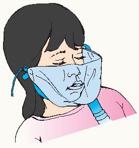

    Oxygen by mask


    1. Choose an appropriate-size mask that covers the mouth and nose of the child but not the eyes.


    2. Use a mask that is capable of delivering the desired oxygen concentration.


    3. Place the mask over the child’s mouth and nose so it fits securely. Secure the mask with an elastic head grip.


    4. Remove the oxygen mask at hourly intervals; wash and dry the face.


    5. Do not use masks for comatose infants or children.


    6. For additional information, refer to Procedure Guidelines 10-14, page 242, and Procedure Guidelines 10-13, page 240.


    1. Extra space under mask and around face is added dead space and decreases effectiveness of the therapy.


    2. Venturi masks for pediatric use deliver low to moderate concentrations of oxygen: 24%, 28%, 31%, 35%, or 40%.


    3. Make sure the mask is adjusted properly over the mouth and nose. Do not allow the oxygen to blow in the child’s eyes.


    4. Makes the patient feel more comfortable.


    5. Such children are more likely to vomit. The risk of aspiration may be increased with mask therapy because of obstruction of the flow of vomitus.


    Face tent


    1. Face tents are available in adult size only. They can be used effectively in pediatric patients if inverted to create a smaller reservoir and better fit.


    2. A flow of 8-10 L/minute should be used to flush the system and provide a stable oxygen concentration.


    1. Face tents combine the positive qualities of aerosol masks and mist tents. The child is accessible and may continue to play without feeling confined.


    2. Larger children will require higher flows.


    imageA child receiving humidity and oxygen by way of an aerosol face tent.


    T-Bars and tracheostomy masks


    1. These devices are used to deliver oxygen to intubated patients.


    2. The flow rate must be set to meet the minute volume requirements of the child and to provide a 100% source of gas.


    2. T-bars require a short, flexible tube on the distal end to act as a reservoir and prevent room-air entrapment.


    Closed incubator and Isolettes


    1. The incubator is used to provide a controlled environment for the neonate.


    2. Adjust the oxygen flow to achieve the desired oxygen concentration.


      1. An oxygen limiter prevents the oxygen concentration inside the incubator from exceeding 40%.


      2. Higher concentrations (up to 85%) may be obtained by placing the red reminder flag in the vertical position.


    3. Secure a nebulizer to the inside wall of the incubator if mist therapy is desired.


    4. Keep sleeves of incubator closed to prevent loss of oxygen.


    5. Periodically analyze the incubator atmosphere.


    6. Drain and fill reservoirs with sterile water at least every 24 hours.


    1. The unit is able to provide precise environmental control of temperature, oxygen, humidity, and isolation.


    2. Follow manufacturer’s instructions for each make of incubator.


      1. This is desirable because it reduces the hazard of the child’s developing retinopathy of prematurity.


      2. This operates by reducing the air intake.


    3. This should be cleaned and autoclaved daily. Sterile solutions are used to keep the bacteria count at a minimum.


    4. When the incubator or sleeves are opened, supply supplemental oxygen with oxygen mask to face and nose.


    5. Be certain the child is receiving the desired concentration of oxygen.


    6. Decreases risk of Pseudomonas contamination.


    Oxygen hood/box


    1. Warmed, humidified oxygen is supplied through a plastic container that fits over the child’s head. This method is more suitable for a child under 1 year of age. image


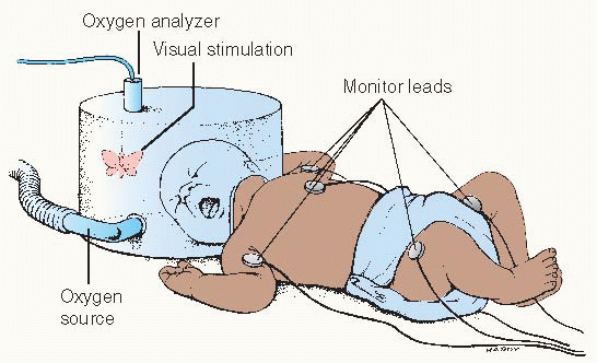
    2. Continuously monitor the oxygen concentration, temperature, and humidity inside the hood.


    3. Open the hood or remove the baby from it as infrequently as possible.


    4. Several different designs are available for use. The manufacturer’s directions should be carefully followed.


    1. This is especially useful when high concentrations of oxygen are desired. The hood may be used in an incubator or with a warming unit.


      Oxygen should not be allowed to blow directly into the infant’s face.


    2. Oxygen should be warmed to 87.8°-93.2° F (31°-34° C) to prevent a neonatal response to cold stress, including oxygen deprivation, metabolic acidosis, rapid depletion of glycogen stores, and reduction of blood glucose levels.


    3. Prevents fluctuations of heat and oxygen, which may further debilitate the young infant.


    4. This is a safety consideration.


    image Evidence Base Ghuman, A. K., Newth, C. J. L., & Khemani, R. G. (2010). Respiratory support in children. Paediatrics and Child Health, 21, 4.


  • Nursing care should continue to address issues of hydration, nutrition, sedation, skin integrity, tissue perfusion, infection control, communication, safety, and parental support and education.


  • Available ventilators for pediatric use have a wide range of capabilities, versatility, and clinical application. Some are more suitable for use with infants, others with older children.


    • Nurses must be well acquainted with the characteristics of the particular machine being used and the meaning of the settings and alarms on the machine.


Nursing Management

See Procedure Guidelines 10-19, page 254. In addition, the nurse who is caring for a pediatric patient should remember the following.


Setting Controls

In setting controls, inspiratory flow rate will be less and the respiratory rate will be greater than in the adult patient. These depend on the patient’s size and condition and are determined by the health care provider or respiratory therapist.


Humidification

During ventilation of an infant in an incubator, the amount of ventilator tubing outside the incubator should be kept to a minimum. The warm temperature inside the incubator helps decrease the amount of condensation in the tubing and thus provides higher water content in the inspired gas.


Oxygen Concentration


  • In infants, inspired concentrations of oxygen should always be kept as low as possible (while still providing for physiologic requirements) to prevent the development of retinopathy of prematurity or pulmonary oxygen toxicity.


  • The oxygen concentration should be checked periodically with an analyzer.


Blood Gases


  • Blood gas analysis to monitor oxygenation, via arteriopuncture or umbilical or arterial lines.


  • The arterialized capillary sample method is inaccurate for infants in respiratory distress because the constricted peripheral circulation may not reflect the ABG (arterial blood gases) levels accurately.


Sterile Precautions

The neonate has only those antibodies transferred across the placenta from the mother. Therefore, sterile precautions are essential.


  • Ventilator tubing should be changed every 24 hours.


  • Routine cultures should be taken after intubation; there should be daily Gram staining of secretions.


  • Suctioning requires aseptic technique.


Tubing Support


  • Special frames are available to support ventilator tubing; this helps to prevent accidental decannulation in infants and small children.


  • Infants may require support or padding on either side and at the top of their heads to decrease mobility and take up space between the head and the frame.


Monitoring the Ventilator


  • Pressure gauges should be checked at frequent intervals because this gives an indication of changing compliance or increased airway resistance.


  • Volume measurements are difficult to obtain in infants because most spirometers incorporated into ventilators and meters do not read accurately at low volumes and flows. However, they are helpful with older children.


  • Measure respiratory rates of the machine and the patient at least every hour and record.


DISORDERS


Common Pediatric Respiratory Infections

Respiratory tract infection is a frequent cause of acute illness in infants and children. Many pediatric infections are seasonal. The child’s response to the infection will vary based on the age of the child, causative organism, general health of the child, existence of chronic medical conditions, and degree of contact with other children. Information about specific respiratory infections, including bacterial pneumonia, viral pneumonia, Pneumocystis pneumonia, Mycoplasma pneumonia, bronchiolitis, croup, and epiglottitis, may be found in Figure 44-1 and Table 44-1, pages 1484 to 1492.


Nursing Assessment

Determine the severity of the respiratory distress that the child is experiencing. Make an initial nursing assessment.


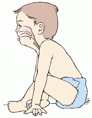
Figure 44-1. Characteristic posture of a child with acute epiglottitis: leaning forward on hands, in the “tripod” position; mouth open, tongue out, head forward and tilted up in a sniffing position in an effort to relieve the acute airway obstruction secondary to swollen epiglottis.


Table 44-1 Common Pediatric Respiratory Infections


CONDITION AND CAUSATIVE AGENT


AGE AND INCIDENCE


CLINICAL MANIFESTATIONS


DIAGNOSTIC EVALUATION


TREATMENT


NURSING CONSIDERATIONS


COMPLICATIONS


Bacterial Pneumonia


Bacterial infection of the lung parenchyma.


General considerations: In the normal child, bacterial pneumonias are not common. A viral respiratory infection commonly occurs before bacterial pneumonia. The initial viral infection alters the lungs’ defense mechanisms.


Pneumococcal pneumonia


• The most common causative agent is Streptococcus pneumoniae.


image NURSING ALERT Pneumococcal polysaccharide vaccine provides protection from 23 types of Streptococcus pneumoniae. It is recommended for children age 2 years and older with sickle cell disease, functional or anatomic asplenia, nephrotic syndrome, human immunodeficiency virus (HIV) infection, and Hodgkin’s disease before beginning cytoreduction therapy.


• Responsible for the majority of bacterial pneumonias in ages 1 month through 6 years. However, it is seen in all age groups.


• The incidence has declined due to the use of the vaccine.


• Most common in winter and spring.


Infants: Mild URI of several days’ duration, poor feeding, decreased appetite.


• Abrupt onset of fever 102.2° F (39° C) or higher; restlessness, respiratory distress, air hunger, pallor, cyanosis (common), nasal flaring, retractions, grunting, tachypnea, tachycardia, irritability; may see abdominal distention due to swallowed air or ileus.


Older child: Mild URI, followed by fever up to 104.9° F (40.5° C), shaking chills, headache, decreased appetite, abdominal pain, vomiting, drowsiness, restlessness, irritability, lethargy, rhonchi, fine crackles, dry hacking cough, increased respirations, anxiety, occasionally circumoral cyanosis, pleuritic pain, diminished breath sounds, may develop a pleural effusion, empyema.


• Chest x-ray: Commonly does not correspond to clinical findings.


Infants: Patchy, diffuse areas follow a bronchial distribution, many limited areas of consolidation around smaller airways.


Young and older child: Lobar or segmental consolidation; pleural fluid may be present.


• WBC count elevated, ABG analysis indicates hypoxemia.


Cultures: Sputum, nasopharyngeal secretions, pleural fluid, blood.


• Oxygen.


• Penicillin G; penicillin allergy: erythromycin, trimethoprim-sulfamethoxazole, alternately amoxicillin, ampicillin, cefuroxime, cefotaxime, ceftriaxone, clindamycin, chloramphenicol.


• Due to the increased incidence of penicillinaseresistant pneumococci, all pneumococcal isolates should be tested for resistance. Vancomycin has been recommended for penicillin-resistant strains.


• Bronchodilators.


• Bed rest, monitor fluids, intake and output. Give oral fluids cautiously to avoid aspiration. Do not give oral fluids to a child in respiratory distress.


• Administer oxygen with humidification. Perform frequent, thorough respiratory assessment.


• Administer antipyretics as prescribed.


• Change position frequently. Isolation procedures, as ordered or per facility policy.


• Rare, but include bacteremia, empyema, pleural effusion, otitis media, sinusitis, meningitis, hemolytic uremic syndrome, lung abscess, necrotizing pneumonia.


Streptococcal pneumonia


• Beta-hemolytic Streptococcus group A


• Ages 3-5 years. Uncommon, serious.


• An endemic influenza predisposes to streptococcal pneumonia and tracheobronchitis.


• Sudden onset, high fever, chills, worsening cough, pleuritic pain, respiratory distress, grunting, retractions, altered mental status, signs of shock, decreased capillary refill, tachycardia.


• May be insidious, mildly ill, low-grade fever.


• Severe infections—toxic shock syndrome with erythematous rash, desquamation, hypotension, hepatic dysfunction, renal involvement, vomiting and diarrhea, hematologic abnormalities, acute respiratory distress syndrome.


• Initial chest x-ray may be normal or slightly abnormal. Within 24 hours, chest x-ray worsens.


• Unilateral lobar disease, bilateral diffuse infiltrates with severe disease.


• Blood cultures, nasopharyngeal secretions cultured, throat swab, culture pleural fluid and lung aspirate.


• Leukocytosis, sedimentation rate increased, elevated serum ASO titer.


• Penicillin G


• Administer antipyretics.


• Provide humidified oxygen, as needed. Rest. Monitor intake and output.


• Empyema, toxic shock syndrome, severe respiratory compromise, pneumatoceles. Can be life-threatening.


Staphylococcal pneumonia


• Staphylococcus aureus, Gram-positive


Most common in children ages 6 months to 1 year. October-May (rare)


• Predisposing factors: cystic fibrosis, maternal infection, immunodeficiency.


• Usually preceded by a viral URI. Changes abruptly to high fever, cough, respiratory distress, tachypnea, grunting, nasal flaring, cyanosis, retractions.


• Anxiety, lethargic, occasionally vomiting, diarrhea, anorexia, abdominal distention, toxic appearance.


Older infant/child: Elevated WBC count, especially polymorphonuclear cells.


Young infant: WBC count may be normal, mild to moderate anemia.


• Cultures—pleural fluid, lung aspirate, sputum, gastric aspirate, blood.


• If pulmonary fluid is purulent and of a large amount, closed chest drainage may be utilized.


• Chest x-ray: Patchy infiltrate, may involve entire lobe or hemithorax. Right lung involvement common; bilateral involvement is also seen. Pleural effusion, empyema, pyopneumothorax, pneumatoceles, pneumothorax, lung abscesses.


• Thoracentesis


• Nafcillin, oxacillin, methicillin, cefazolin, clindamycin, vancomycin. MRSA exists, especially in long-term care facilities or in patients with a prolonged hospital stay.


• Isolation per policy, check for MRSA, prevent nosocomial infection.


• Rapid treatment is important. Administer antibiotics as soon as possible. Monitor for signs of tension pneumothorax. Monitor fluid status closely.


• Strict handwashing.


• Staphylococcal pneumonia is rare and must be treated aggressively due to rapid onset and deterioration.


• Empyema, tension pneumothorax, abscess, fibrothorax, bronchiectasis, osteomyelitis, staphylococcal pericarditis. Consider screening infants for cystic fibrosis and immunodeficiency.


Haemophilus influenzae, type B


• Majority of children younger than age 4 years.


• Infants and children who are not immunized.


• Winter and spring.


• Usually preceded by URI. Associated with otitis media, epiglottitis, and meningitis; appears toxic.


• Insidious onset; cough, febrile, tachypneic, nasal flaring, retractions.


• Chest x-ray: Usually lobar infiltrates; however, segmental, single, or multiple lobe infiltrates are also seen. Pleural effusion, pneumatocele. CBC: elevated WBC, lymphopenia.


• Cultures: blood, pleural fluid, lung aspirates, and nasal secretions. In the absence of a positive culture, a positive urine latex agglutination can confirm diagnosis.


• If atelectasis is present, bronchoscopy to rule out foreign body.


• Patients may present receiving antibiotics for otitis media.


• Ceftriaxone and other cephalosporins, ampicillin, chloramphenicol, azithromycin.


• Administer antibiotics on time.


• Ensure adequate hydration. Monitor for signs of upper respiratory impairment, drooling, stridor, and dusky color.


• Respiratory isolation until 24 hours after appropriate antibiotic therapy is initiated.


• Rifampin prophylaxis should be considered for close household contacts if there are:


• Incomplete or unvaccinated members younger than age 2 years.


• Immunocompromised child.


• Daycare setting with two or more cases of invasive disease within 2 months and incompletely vaccinated children in attendance.


• Not recommended for pregnant women.


• Frequently in young infants, bacteremia, pericarditis, cellulitis, empyema, meningitis, pyarthrosis.


Viral Pneumonia


• Respiratory syncytial virus (RSV); parainfluenza virus types 1, 2, 3; Adenoviruses types 1, 2, 5, 6 and types 3, 7, 11, 21; Influenza A and B.


• Peak age for bronchiolitis is within the first year of life. Peak age for viral pneumonia is age 2.3 years.


• Typically seen in the winter months.


• Usually preceded by URI with symptoms of cough, rhinitis, and mild fever. Progressing to tachypnea, poor feeding in infants and retractions (suprasternal, intercostal, subcostal and substernal), leading to nasal flaring. Along with use of accessory muscles, wheezing, severe cough, cyanosis, and respiratory fatigue.


• Viral pneumonia may present concurrently with bacterial pneumonia. Adenovirus types 3, 7, 11, and 21 may cause severe necrotizing pneumonia in infants.


• Chest x-ray shows patchy infiltrates, transient lobal infiltration, and hyperinflation.


• CBC: slightly elevated WBC count.


• Cultures: blood and nasopharyngeal secretions.


• Viral antigens for rapid diagnosis.


• If bacterial pneumonia is suspected, administer antibiotics.


• Supportive measures: IV fluids, antipyretics, humidified oxygen, assisted ventilation.


• Avoid aspirin due to risk of Reye’s syndrome.


• Oseltamivir or zanamivir for influenza infection.


• Monitor closely for signs of respiratory fatigue or distress.


• Monitor oxygen saturation levels and response to oxygen therapy if hypoxic.


• Close cardiac monitoring may be indicated.


• Monitor for adequate hydration and nutritional status. Elevate infants up to an angle of 10-30 degrees to ease breathing. Infants and children who require mechanical ventilation require close supervision and frequent monitoring. Institute contact precautions with strict handwashing.


• Prevention: influenza vaccine.


• Influenza: severe fulminant pneumonia with hemorrhagic exudate. Death may result. Severe disease may be seen in children with cardiopulmonary disease, cystic fibrosis, bronchopulmonary dysplasia, and neurovascular disease.


• Type B—myositis.


• RSV subgroup A (more virulent, associated with more severe disease); RSV subgroup B


• In infants, RSV is the most common cause of pneumonia, bronchiolitis, and hospitalizations.


• Severity of RSV infection decreases with age and subsequent infections.


• Peak age is 2-7 months, October-April.


• Typically begins with URI, rhinorrhea, fever usually less than 102° F (38.9° C), otitis media, and conjunctivitis.


• Progressing to coughing, wheezing, tachypnea (greater than 70 breaths/minute), intercostal and subcostal retractions, poor air exchange, hypoxia, decreased breath sounds, cyanosis, lethargy, listless apneic episodes, and irritability. Infants will present with poor feeding, inability to suck and breathe.


• Nasopharyngeal secretions for rapid antibody or assay for RSV antigen detection.


• Chest x-ray: chest hyperexpanded, air trapping, multiple lobe infiltrates, atelectasis.


• ABG analysis.


• Pulse oximetry.


• Supportive measures, such as oxygen, respiratory support, hydration.


• Bronchodilators should not be used routinely.


• Racemic epinephrine can be effective in bronchiolitis.


• Glucocorticoids and ribavirin are not supported in the literature.


• Antibiotics only if a secondary bacterial infection is suspected.


• Early detection is important in hospitalized patients. Institute contact precautions with strict handwashing. RSV Immune Globulin (RSV-IGIV) and palivizumab may be given to prevent infection in select patient populations. Consult AAP Guidelines for specific information.


• Prevent nosocomial spread. Institute contact isolation. Strict handwashing.


• RSV-positive patients should not be in contact with other high-risk patients, such as those with chronic cardiac or respiratory illness or immunocompromised patients.


• Assess frequently for signs of respiratory failure.


• Use noninvasive oxygen monitoring.


• In tachypneic patients and those in respiratory distress, oral fluids are contraindicated due to risk of aspiration.


• RSV can be fatal, especially in children with chronic cardiac and respiratory diseases, premature infants, and those with underlying neuromuscular or immunologic diseases.


• Respiratory failure, intubation, and mechanical ventilation.


• In older children with asthma, RSV can cause an acute asthmatic episode.


Pneumocystis Carinii Pneumonia


• P. carinii, also known as P. jiroveci, is a fungus with similarities to a protozoa. The organism exists in three forms in the tissues: trophozoite, sporozoite, and cyst.


• Most healthy humans are infected before age 4 years and are asymptomatic.


• Life-threatening pneumonia is seen in the immunosuppressed host. PCP in severely immunocompromised children with acquired or congenital immunodeficiency disorders, malignancies, organ transplant recipients, and debilitated, malnourished, and premature infants. In HIV-infected children it most commonly occurs between ages 3 and 6 months. PCP can occur during remission or relapse in patients with leukemia or lymphoma.


• Slow onset, tachypnea, retractions, and nasal flaring cyanosis.


• Sporadic form in immunocompromised patients. Signs may vary; onset may be acute or fulminant.


• Fever, tachypnea, dyspnea, cough, nasal flaring, cyanosis, subacute, diffuse pneumonitis with dyspnea at rest, tachypnea and decreasing oxygen saturation. Extrapulmonary sites rarely occur and usually produce no symptoms.


• Chest x-ray: Bilateral, diffuse, alveolar disease with a granular pattern, initially perihilar densities, which progress to peripheral and apical areas.


• Organism cannot be cultured from routine specimens.


• Open lung biopsy is the most reliable method.


• Bronchoalveolar lavage is also utilized to obtain samples. Needle aspiration of the lung. IgM-ELISA: elevated. CBC: mild leukocytosis, moderate eosinophilia.


• PCP mortality is 5%-40% in immunocompromised patients; if untreated, 100%.


• Trimethoprim-sulfamethoxazole; rate of adverse reactions is high in HIV-infected patients.


• Pentamidine, parenterally or aerosolized. IV form associated with increased risk of adverse reactions. Aerosolized for children ages 5 and older.


• Corticosteroids recommended for children older than age 13, may be used in younger children.


• Close monitoring of respiratory status, hydration, and nutrition.


• Monitor for adverse reactions to therapy: vomiting, nausea, rash.


• Respiratory isolation should be instituted for 48 hours after initiation of therapy.


• PCP prophylaxis (see page 1717).


image NURSING ALERT Pentamidine is associated with a high incidence of adverse reactions: pancreatitis, renal dysfunction, hypoglycemia, hyperglycemia, hypotension, fever, and neutropenia. Pentamidine should not be used with didanosine, which also causes pancreatitis, and in patients with hepatic dysfunction.


Mycoplasma Pneumonia


Mycoplasma pneumoniae, microorganisms with properties between bacteria and viruses.


• Fall and winter.


• Crowded living conditions. Seen frequently in school age children and adolescents.


• Slow onset; 2- to 3-week incubation period. Coryza, malaise, headache, anorexia, normal temp or low-grade fever, sore throat, muscle pain, vomiting, subacute tracheobronchitis, shortness of breath, dry cough that progresses to mucopurulent cough, mild chest pain, wheezing. May present with maculopapular rash.


• Chest x-ray: bronchopneumonic, diffuse bilateral infiltrates.


• Complement fixation test: increased. Cold agglutinins: increased. Immunofluorescent and enzyme immunoassay tests.


• Positive sputum culture.


• Erythromycin, azithromycin, clarithromycin, tetracycline, and doxycycline may be used in children ages 8 and older.


• Children should be on secretion precautions.


• Monitor fever.


• Assess need for cough suppressants.


• May be fatal if infection becomes systemic or if the child has a preexisting chronic lung disease, sickle cell anemia, immunodeficiencies, or cardiac disease. Those with Down syndrome can develop severe pneumonia.


• Pleural effusions.


Bronchiolitis


• Inflammation of the bronchioles. Causative agents: RSV, adenovirus, parainfluenza type 1 or 3, influenza virus, and M. pneumoniae.


• Winter and spring.


• Most common in infants younger than 6 months old, may occur up to age 2 years.


• Greater incidence in males than females.


• Increased in daycare centers.


• Gradual onset after exposure to an individual with URI. Coryza, tachypnea, respiratory rate greater than 50 bpm, retractions, wheezing, paroxysmal cough, fever, cyanosis, dehydration, poor feeding, vomiting, tachycardia, irritability, dyspnea.


• Apnea may be first sign in infants with RSV. Decreased breath sounds with prolonged expiratory phase. Hypoxemia may persist for 4-6 weeks.


• Chest x-ray: patchy or peribronchial infiltrates, hyperinflation of lungs with flattening of diaphragms. Some will present with a normal chest x-ray.


• Nasopharyngeal viral cultures.


• Serologic studies for specific organisms. ABG analysis for children with respiratory distress.


• Humidified oxygen, ventilatory assistance as needed. (Note treatment for RSV above regarding bronchodilators, glucocorticoids, antibiotics.)


• Antipyretics for fever.


• Continuous cardiac monitoring for apnea or bradycardia.


• Pulse oximetry.


• Avoid high-density humidity; may cause bronchospasm.


• Monitor fluid and electrolyte balance closely. Place infant on apnea monitor.


• Keep nasal passages free of secretions; infants are obligate nose breathers.


• Position patient upright to facilitate breathing. Monitor closely for signs of impending respiratory failure.


• There is a high risk of cross-contamination to noninfected children. Institute contact precautions and respiratory isolation.


• Monitor oxygenation levels with noninvasive monitoring.


• Increasing respiratory distress resulting in the need for mechanical ventilation.


• Secondary bacterial infection.


• Pneumothorax and pneumomediastinum. Apneic episodes, may be lifethreatening in children with chronic respiratory or cardiac disease.


• Some infants demonstrate abnormal lung functions months after infection.


Croup Syndromes


Croup syndromes refer to infections of the supraglottis, glottis, subglottis, and trachea.


Acute laryngotracheobronchitis (subglottic croup)


• Parainfluenza types 1, 2, 3; RSV; influenza A and B; adenovirus; measles.


image NURSING ALERT If ABG analysis is done, a normal partial pressure of arterial carbon dioxide may not indicate decreased severity. By the time hypercapnia is seen, intubation will be required.


• Ages 3 months to 5 years. Peak ages 1-2 years.


• Greater incidence in males than females.


• Late autumn, early winter.


• Usually a preceding URI is seen. Initially, a mild brassy or barking cough, hoarse, intermittent stridor, progresses to continuous stridor.


• Nasal flaring, suprasternal, infrasternal, intercostals, retractions.


• Breathing labored, prolonged expiratory phase. Temperature slightly elevated.


• Crying and agitation aggravate signs.


• Child prefers to be held upright or sit up in bed. Symptoms are worse at night.


• Severe croup: restlessness, air hunger, decreased breath sounds, hypoxemia, hypercapnia, anxiety, cyanosis, tachycardia, and cessation of breathing.


• Diagnosis is made by clinical evaluation and careful history. A croup score may be assigned to grade severity. Lateral neck x-ray: subglottic edema, narrowing with normal supraglottic structures. Anterior/posterior neck film: steeple sign.


• Lower airway involvement.


• Racemic epinephrine nebulized with oxygen. Monitor pulse and cardiac rhythm.


• Severe airway edema may require intubation.


• Corticosteroid therapy.


• Antibiotic therapy.


• Maintain a calm environment, avoid agitating the child, and disturb as little as possible. Monitor oxygenation with noninvasive pulse oximeter. Monitor respiratory status closely and frequently. Closely observe response to racemic epinephrine. Have intubation equipment at bedside and available during transport. Increasing tachypnea may be first sign of hypoxia. If severe distress does not respond to initial treatment, ABG analysis should be obtained. In a hypoxic, pale, cyanotic, or obtunded patient, do not manipulate larynx and do not examine with a tongue blade; it may lead to sudden cardiopulmonary arrest.


• Dehydration, intubation, airway obstruction, death.


Spasmodic croup and acute spasmodic laryngitis


• No infectious agents seen. Thought to be related to spasm of laryngeal muscle.


• Cause may be viral in a few cases, allergic or psychological.


• Ages 1-6 years.


• Peak incidence 7-36 months.


• Similar to acute laryngotracheobronchitis. Symptoms are sudden, in the evening.


• Afebrile, barking, brassy cough, hoarse, stridor. Child may be anxious and frightened.


• Breathing is noisy on inspiration, slow, and labored; respiratory distresses.


• Tachycardia. Skin cool, moist. Dyspnea is worse with excitement.


• Cyanosis is rare; severity decreases over time. Patient appears well in the morning, may cough or sound hoarse. The symptoms may recur for subsequent nights; however, they will be less severe.


• CBC is normal, x-ray: subglottic narrowing.


• Endoscopic exam: inflammation of the arytenoids cartilage, epithelium intact, pale mucosa.


• Corticosteroid therapy.


• Racemic epinephrine, nebulized.


• Same as for acute laryngotracheobronchitis (above).


• Allow the parent to hold the child, remain in upright position.


• Use racemic epinephrine with caution and provide cardiac monitoring, due to possible tachycardia.


Bacterial tracheitis (pseudomembranous croup)


• Tracheal inflammation in the subglottic region.


S. aureus, H. influenzae, streptococci groups A and B, Escherichia coli, Klebsiella, Moraxella catarrhalis, Pseudomonas, Chlamydia trachomatis, and Corynebacterium diphtheriae should be considered in the nonvaccinated patient.


• No season variation.


• Age is variable, 1 month to 6 years; however, most are younger than age 3 years.


• Usually follows a viral illness. Slow or sudden deterioration, child appears toxic.


• High fever, stridor, hoarse, respiratory distress. Thick, purulent, copious airway secretions.


• Mucosal necrosis, brassy or barking cough. Epiglottitis may coexist. May cause life-threatening airway obstruction.


• Neck x-ray: subglottic narrowing, large epiglottis, thick arytenoepiglottic folds, pseudomembrane in trachea.


• Tracheal culture. Laryngoscopy, CBC, leukocytosis, bandemia.


• Humidified oxygen as required, mist, antibiotic therapy, cephalosporin, antipyretics.


• Admit to the intensive care unit (ICU), intubation, and frequent suctioning.


• Racemic epinephrine ineffective.


• Monitor closely for airway obstruction.


• Airway obstruction, death, tracheostomy, pneumothorax, toxic shock syndrome.


Acute epiglottitis


• Supraglottitis, inflammation of the epiglottis and edema of the arytenoepiglottic folds.


H. influenzae type B, most common. S. pneumoniae, S. aureus, group A. Beta-hemolytic streptococcus, Streptococcus pyogenes, Moraxella catarrhalis, and Candida albicans (in the immunocompromised).


• Range ages 2-7 years; peak ages 3-5 years.


• Autumn and winter.


• Incidence significantly decreased due to the routine use of the H. influenzae vaccine.


• Sudden fulminating course, high fever, toxic appearance, sore throat, drooling, dysphagia, aphonia, retractions, air hunger, anxiety, tachycardia, hoarseness, irritability, restlessness, rapid progression to respiratory distress.


• Sitting forward, neck hyperextended, mouth open, tongue protruding. Older child will sit in tripod position. Drooling, agitation, and absence of spontaneous cough are predictive of epiglottitis.


• Absence of croupy cough.


• Complete fatal airway obstruction and death may occur within hours if not treated. In group A beta-hemolytic streptococcus and H. influenzae, patient will present with acute respiratory distress.


• Stridor and breath sounds decrease as child begins to tire.


• A brief episode of air hunger with restlessness and agitation may rapidly progress into increasing cyanosis, coma, and death.


• Clinical evaluation and history, determining the onset of symptoms.


• Monitor oxygen saturation level.


• Lateral neck films: while sitting in parent’s lap with portable radiology, swollen epiglottis.


• Direct exam or laryngoscopy: large, swollen cherry-red epiglottis, edema of arytenoepiglottic folds. CBC, cultures, IV catheters must be done after intubation.


• Medical emergency.


• Establish a stable artificial airway first. Approach child in a calm manner; emotional upset and agitation may result in complete airway obstruction.


• Proceed to the operating room or ICU with personnel skilled and equipped to intubate or perform a percutaneous tracheostomy.


• Lateral neck films should be done in the operating room or ICU after airway is established.


• Antibiotics: intravenous cefotaxime, ceftriaxone, ampicillin with sulbactam.


• Supplemental humidified oxygen; mechanical ventilation, if necessary.


• If epiglottitis is strongly suspected, examination of the throat is contraindicated; due to reflex laryngospasm, acute airway obstruction, aspiration, and cardiopulmonary arrest during or immediately after examination of the pharynx with a tongue blade. Do not attempt throat culture.


• Allow the parents to remain with and hold the child. Before transport to the operating room, observe closely for signs of airway obstruction. Allow the child to maintain a position of comfort (not supine).


• Equipment for intubation and tracheostomy must remain with the patient at all times. A practitioner skilled in intubation and tracheostomy procedures must accompany the child to the operating room or ICU. After intubation, the child should remain in the ICU with frequent assessment of oxygenation levels and need for mechanical ventilation. Prevent self-extubation; use arm boards or restraints to prevent arm movements, extubation, and death. After intubation, administer sedation, as needed. When the decision is made to extubate the child, emergency tracheostomy and intubation equipment must be at bedside.


• Airway obstruction, death, tracheostomy, pneumothorax, toxic shock syndrome.


image NURSING ALERT Do not attempt to visualize the epiglottis with a tongue blade or take a throat culture. May cause laryngospasm and airway obstruction.


ABG, arterial blood gas; ASO, antistreptolysin; CBC, complete blood count; HIV, human immunodeficiency virus; ICU, intensive care unit; MRSA: Methiciííin-resistant S. aureus; PCP, Pneumocystis carinii pneumonia; RSV, respiratory syncytial virus; URI, upper respiratory infection.


  • Observe the respiratory rate and pattern. Count the respirations for 1 full minute, document and note level of activity, such as awake or asleep. Determine if the rate is appropriate for age (see page 1388).


  • Observe respiratory rhythm and depth. Rhythm is described as regular, irregular, or periodic. Depth is normal, hypopnea or too shallow, hyperpnea or too deep.


  • Auscultate breath sounds for a full cycle of inspiration and expiration over all lung fields. Note airflow and presence of adventitious sounds such as crackles, wheeze, or stridor.


  • Observe degree of respiratory effort—normal, difficult, or labored. Normal breathing is effortless and easy.


  • Document character of dyspnea or labored breathing; continuous, intermittent, worsening, or sudden onset. Note relation to activity, such as rest, exertion, crying, feeding, and association with pain, positioning, or orthopnea.


  • Note presence of additional signs of respiratory distress: nasal flaring, grunting, and retractions. Note location of retractions (see Figure 44-2) and character (mild, moderate, or severe).


  • Observe for head bobbing, usually noted in a sleeping or exhausted infant. The infant is held by caregiver with head supported on the caregiver’s arm at the suboccipital area. The head bobs forward with each inspiration.


  • Observe the child’s color. Note the presence and location of cyanosis—peripheral, perioral, facial, and trunk. Note degree of color changes, duration, and association with activity such as crying, feeding, and sleeping.


  • Observe the presence of cough, noting type and duration, such as dry, barking, paroxysmal, or productive. Note any pattern, such as time of day or night, association with activity, physical exertion, or feeding. Severity of croup may be determined by cough and signs of respiratory effort.


    • Mild croup—occasional barking cough, no audible stridor at rest, and either mild or no suprasternal or intercostal retractions.


    • Moderate croup—frequent barking cough, easily audible stridor at rest, and suprasternal and sternal retractions at rest, but little or no agitation.


    • Severe croup—frequent barking cough, prominent inspiratory and occasional expiratory stridor, marked sternal retractions, and agitation and distress.


    • Impending respiratory failure—barking cough (often not prominent), audible stridor at rest (may be hard to hear), sternal retractions (may be marked), lethargy or decreased level of consciousness, and often dusky appearance in the absence of supplemental oxygen.

      image Evidence Base
      Ledoux, M., Perkin, R., Sharieff, G. (2012). Upper airway obstruction in pediatrics. Pediatric Emergency Medicine Reports, 17(1), 1-11.


  • Note the presence of sputum, including color, amount, consistency, and frequency.


  • Observe the child’s fingernails and toenails for cyanosis and the presence and degree of clubbing, which indicate underlying chronic respiratory disease.


  • Evaluate the child’s degree of restlessness, apprehension, and muscle tone.


  • Note the presence or complaint of chest pain and its location, whether it is local or generalized, dull or sharp, and associated with respiration or grunting.


  • Assess for signs of infection, such as elevated temperature; enlarged cervical lymph glands; purulent discharge from nose or ears; sputum; or inflamed mucous membranes.


Figure 44-2. Sites of respiratory retractions.


Nursing Diagnoses


  • Ineffective Airway Clearance related to inflammation, obstruction, secretions, or pain.


  • Ineffective Breathing Pattern related to inflammatory process or pain.


  • Deficient Fluid Volume related to fever, decreased appetite, and vomiting.


  • Fatigue related to increased work of breathing.


  • Anxiety related to respiratory distress and hospitalization.


  • Parental Role Conflict related to hospitalization of the child.


Nursing Interventions


Promoting Effective Airway Clearance


  • Provide a humidified environment enriched with oxygen to combat hypoxia and to liquify secretions (see Procedure Guidelines 44-1, pages 1480 to 1482).


  • Advise the parents to use a jet or ultrasonic nebulizer at home and encourage fluids, as tolerated.


  • Keep nasal passages free of secretions. Infants are obligate nose breathers. Use a bulb syringe to clear nares and oropharynx.


Improving Breathing Pattern


  • Place the child in a comfortable position to promote easier ventilation.


    • Semi-Fowler’s—use infant seat or elevate head of bed.


    • Occasional side or abdominal position will aid drainage of liquefied secretions. Do not place infant in prone position.


    • Do not position the child in severe respiratory distress in a supine position. Allow the child to assume a position of comfort.

      image NURSING ALERT To minimize spasm and sudden blockage of airway, avoid the following: making the child lie flat, forcing the child to drink, and looking down the child’s throat.


  • Provide measures to improve ventilation of the affected portion of the lung.


    • Change position frequently.


    • Provide postural drainage, if prescribed.


    • Relieve nasal obstruction that contributes to breathing difficulty. Instill normal saline solution or prescribed nose drops and apply nasal suctioning.


    • Quiet prolonged crying, which can irritate the airway, by soothing the child; however, crying may be an effective way to inflate the lungs.


    • Realize that coughing is a normal tracheobronchial cleansing procedure, but temporarily relieve coughing by allowing the child to sip water; use extreme caution to prevent aspiration.


    • Insert a nasogastric tube, as ordered, to relieve abdominal distention, which can limit diaphragmatic excursion.


  • Ensure that the child’s oxygen input is not compromised.


    • Monitor oxygen saturations, as indicated; pulse oximetry should be performed if hypoxia is suspected.


    • If compressed air or oxygen is administered, use a head box to avoid excess carbon dioxide concentrations and increased respiratory rate.


  • Administer appropriate antibiotic or antiviral therapy.


    • Observe for drug sensitivity.


    • Observe the child’s response to therapy.


  • Administer specific treatment for respiratory syncytial virus (RSV), if ordered.


    • There is presently no evidence that supports the use of ribavirin or inhaled corticosteroids in acute bronchiolitis; however, some health care providers do consider these options in the management of RSV.

      image Evidence Base
      Kelsall-Knight, L. (2012). Clinical assessment of a child with bronchiolitis. Nursing Children & Young People, Oct; 24(8), 29-34.


  • If the decision is made to initiate ribavirin therapy, appropriate precautions should be implemented to protect personnel and caregivers.


    • Information should be provided about the potential but unknown risk of exposure to ribavirin.


    • Pregnant women should be advised to refrain from providing direct care to patients who are receiving ribavirin therapy.


    • Methods to reduce environmental exposure to ribavirin should be employed:


      • Stop aerosol administration before opening hood or tent.


      • Use room with adequate ventilation of at least six air exchanges per hour.


      • Consider the use of scavenger devices to help decrease the escape of ribavirin into the air.


      • Staff should wear aprons and face masks for the duration of the time the nebulizer is in use.


  • For cases of severe respiratory distress, assist with intubation or tracheostomy and mechanical ventilation.


    • Tracheostomy and endotracheal tubes are generally not cuffed for infants and small children because the tube itself is big enough relative to the size of the trachea to act as its own sealer.


    • Position the infant with a tracheostomy with the neck extended by placing a small roll under the shoulders to prevent occlusion of the tube by the chin. Support his or her head and neck carefully when moving the infant to prevent dislodgement of the tube.


    • When feeding, cover the tracheostomy with a moist piece of gauze, or use a bib for older infants or young children.


    • See page 1479 in this chapter as well as Chapter 10 for care of the patient on mechanical ventilation.

      image NURSING ALERT Infants with a history of very low birth weight and bronchopulmonary dysplasia have chronic compensated carbon dioxide retention. Careful attention must be given to oxygen administration to avoid respiratory depression by suppressing their hypoxic drive.


Promoting Adequate Hydration


  • Administer fluids via intravenous (IV) route at the prescribed rate.


  • To prevent aspiration, withhold oral food and fluids if the child is in severe respiratory distress.


  • Offer the child small sips of clear fluid when respiratory status improves.


    • Note any vomiting or abdominal distention after the oral fluid is given.


    • As the child begins to take more fluid by mouth, notify the health care provider and modify the IV fluid rate to prevent fluid overload.


    • Do not force the child to take fluids orally because this may cause increased distress and possibly vomiting. Anorexia will subside as the condition improves.


  • Assist in the control of fever to reduce respiratory rate and fluid loss.


    • Give antipyretics, as prescribed.


  • Record the child’s intake and output and monitor urinespecific gravity.


  • Provide mouth care or offer mouth rinse if child is able to perform this safely.


Promoting Adequate Rest


  • Disturb the child as little as possible by organizing nursing care, and protect the child from unnecessary interruptions.


  • Be aware of the age of the child and be familiar with the level of growth and development as it applies to hospitalization.


  • Encourage the parents to stay with the child as much as possible to provide comfort and security.


  • Provide opportunities for quiet play as the child’s condition improves.


Reducing Anxiety


  • Explain procedures and hospital routine to the child as appropriate for age.


  • Provide a quiet, stress-free environment.


  • Observe the child’s response to the oxygen therapy environment and provide reassurance.


    • The child may experience fear of confinement or suffocation.


    • Vision is distorted through the plastic.


    • The environment is noisy and damp.


    • Physical and diversional activities are restricted.


    • Parental contact is decreased.


    • The environment is often uncomfortable.


  • Avoid the use of sedatives and opiates, which may obscure restlessness. Restlessness is a sign of increasing respiratory distress or obstruction.


  • Allow the child to assume a position of comfort.


Strengthening the Parents’ Role


  • Help the parents understand the purpose of the oxygen therapy/humidifier and how to work with it.


  • Discuss their fears and concerns about the child’s therapy.


  • Include the parents in planning for the child’s care. Promote their participation in caring for the child.


  • Recognize that the parents will need rest periods. Encourage them to take breaks and eat on a regular basis.


Family Education and Health Maintenance


  • Teach the importance of good hygiene. Include information on handwashing and appropriate ways to handle respiratory secretions at home.


  • Teach the family when it is appropriate to keep the child home from school (any fever, coughing up secretions, and significant runny nose in toddler or younger child).


  • Teach methods to keep the ill child well hydrated.


    • Provide small amounts of fluids frequently.


    • Offer clear liquids and prepared electrolyte preparations.


    • Offer frozen juice pops.


    • Avoid juices with a high sugar content.


  • Teach ways to assess the child’s hydration status at home.


    • Decreased number of wet diapers or number of times the child urinates per day.


    • Decreased activity level.


    • Dry lips and mucous membranes.


    • No tears when the child cries.


  • Teach the parents when to contact their health care provider—signs of respiratory distress, recurrent fever, decreased appetite and activity, and signs of dehydration.


  • Teach about medications and follow-up.


  • If a tracheostomy was required, teach care of the tracheostomy, use of equipment, safety, and referral for home nursing care before discharge.


Evaluation: Expected Outcomes


  • Breath sounds clear and equal.


  • Easy, regular, unlabored respirations on room air (or back to baseline if the child is on oxygen).


  • Mucous membranes moist; urine output adequate.


  • Bathing and feeding tolerated well.


  • Child calm and interacts appropriately with family and staff.


  • Parents participate in the child’s care.


Disorders Requiring Surgery of the Tonsils and Adenoids


Tonsillectomy and αdenoidectomy are the surgical removal of the adenoidal and tonsillar structures, part of the lymphoid tissue that encircles the pharynx. These are one of the most frequently performed surgical procedures in the child. The most common disease processes that require tonsillectomy and adenoidectomy are obstructive sleep apnea; chronic, persistent tonsillitis or adenoiditis; and chronic persistent otitis media.


Pathophysiology and Etiology


Function of Tonsils and Adenoids

Jul 20, 2016 | Posted by in NURSING | Comments Off on Pediatric Respiratory Disorders

Full access? Get Clinical Tree

Get Clinical Tree app for offline access