Neurologic Assessment
Neurologic vital signs supplement the routine measurement of temperature, pulse rate, blood pressure, and respirations by evaluating the patient’s level of consciousness (LOC), pupillary activity, and orientation to time, place, and person. They provide a simple, indispensable tool for quickly checking the patient’s neurologic status.
A measure of environmental awareness and self-awareness, LOC reflects cortical function and usually provides the first sign of central nervous system (CNS) deterioration. Changes in pupillary activity (pupil size, shape, equality, and response to light) may signal increased intracranial pressure (ICP) associated with a space-occupying lesion. Evaluating muscle strength and tone, reflexes, and posture also may help identify nervous system damage.
Changes in neurologic vital signs alone rarely indicate neurologic compromise; any changes should be evaluated in light of a complete neurologic assessment. But because these vital signs are controlled at the medullary level, changes in neurologic vital signs may signify ominous neurologic compromise.
Equipment
Penlight ▪ thermometer ▪ sterile cotton ball or cotton-tipped applicator ▪ stethoscope ▪ sphygmomanometer ▪ pupil size chart ▪ pencil or pen.
Implementation
Confirm the patient’s identity using at least two patient identifiers according to your facility’s policy.1
Explain the procedure to the patient, even if he’s unresponsive. Answer all questions to decrease anxiety and increase cooperation.
Assessing LOC and Orientation
Assess the patient’s LOC by evaluating his responses. Use standard methods such as the Glasgow Coma Scale (see Using the Glasgow Coma Scale, page 514) and the Rancho Los Amigos Cognitive Scale (see Using the Rancho Los Amigos Cognitive Scale, page 515). Begin by measuring the patient’s response to verbal, light tactile (touch), or painful (nail bed pressure) stimuli. First, ask the patient his full name. If he responds appropriately, assess his orientation to time, place, and person. Ask him where he is and then what day, season, and year it is. (Expect disorientation to affect the sense of date first, then time, place, caregivers and, finally, self.) When he responds verbally, assess the quality of replies. For example, garbled words indicate difficulty with the motor nerves that govern speech muscles. Rambling responses indicate difficulty with thought processing and organization.
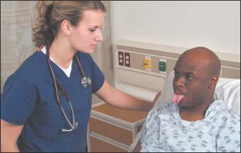
Assess the patient’s ability to understand and follow one-step commands that require a motor response. For example, ask him to open and close his eyes or stick out his tongue (as shown on next column top). Note whether the patient can maintain his LOC. If you must gently shake him to keep him focused on your verbal commands, he may be neurologically compromised.

If the patient doesn’t respond to commands or touch, apply a painful stimulus. Painful stimuli are classified as central (response via the brain) or peripheral (response via the spine). Apply a central stimulus first and note the patient’s response. Acceptable central stimuli include squeezing the trapezius muscle, applying supraorbital or mandibular pressure, and rubbing the sternum. If the patient doesn’t respond to central stimuli, apply a peripheral stimulus to all four extremities to establish a baseline. With moderate pressure, squeeze the nail beds on fingers and toes, and note his response. Check motor responses bilaterally to rule out monoplegia (paralysis of a single area) and hemiplegia (paralysis of one side of the body).
Using the Glasgow Coma Scale
The Glasgow Coma Scale provides a standard reference for assessing or monitoring level of consciousness in a patient with a suspected or confirmed brain injury. This scale measures three responses to stimuli—eye opening, motor response, and verbal response—and assigns a number to each of the possible responses within these categories.
A score of 3 is the lowest and 15, the highest. A score of 7 or less indicates coma. This scale is commonly used in the emergency department, at the scene of an accident, and for evaluation of the hospitalized patient.
| Characteristic | Response/score |
|---|---|
| Eye opening |
|
| Best motor response |
|
| Best verbal response (Arouse patient with painful stimuli, if necessary) |
|
| Total: 3 to 15 | |
Examining Pupils and Eye Movement
Ask the patient to open his eyes. If he doesn’t respond, gently lift his upper eyelids. Inspect each pupil for size and shape, and compare the two for equality (as shown below). To evaluate pupil size more precisely, use a chart showing the various pupil sizes (in increments of 1 mm, with the normal diameter ranging from 2 to 6 mm). Remember, pupil size varies considerably, and some patients have normally unequal pupils (anisocoria). Also see if the pupils are positioned in, or deviated from, the midline. (See Testing the pupils, page 516.)
Stay updated, free articles. Join our Telegram channel

Full access? Get Clinical Tree


Get Clinical Tree app for offline access
