Incentive Spirometry
Incentive spirometry involves using a device to help the patient achieve maximal ventilation by providing feedback on respiratory effort. The device measures respiratory flow or respiratory volume and induces the patient to take a deep breath and hold it for several seconds. This deep breath increases lung volume, boosts alveolar inflation, and promotes venous return. Incentive spirometry is designed to replicate the natural mechanisms of sighing or yawning, thus preventing and reversing the alveolar collapse that causes atelectasis and pneumonitis. It’s suggested
that the patient do 5 to 10 breaths per session every hour while awake (approximately 100 breaths per day).1
that the patient do 5 to 10 breaths per session every hour while awake (approximately 100 breaths per day).1
Devices used for incentive spirometry provide a visual incentive to breathe deeply. Some are activated when the patient inhales a certain volume of air; the device then estimates the amount of air inhaled. Others contain plastic floats, which rise according to the amount of air the patient pulls through the device when he inhales.
Patients at low risk for developing atelectasis may use a flow incentive spirometer. Patients at high risk may need a volume incentive spirometer, which measures lung inflation more precisely.
Incentive spirometry benefits the patient on prolonged bed rest, especially the postoperative patient who may regain his normal respiratory pattern slowly due to such predisposing factors as abdominal or thoracic surgery, advanced age, inactivity, obesity, smoking, or decreased ability to cough effectively and expel lung secretions.
Equipment
Flow or volume incentive spirometer, as indicated, with sterile disposable tube and mouthpiece (the tube and mouthpiece are sterile on first use and clean on subsequent uses) ▪ stethoscope ▪ gloves ▪ watch ▪ pencil and paper ▪ hospital-grade disinfectant ▪ Optional: pillow.
Preparation of Equipment
Gather the ordered equipment at the patient’s bedside. Read the manufacturer’s instructions for spirometer setup and operation. Remove the sterile flow tube and mouthpiece from the package, and attach them to the device. Set the flow rate or volume goal as determined by the doctor or respiratory therapist and based on the patient’s preoperative performance. Turn on the machine if applicable.
Implementation
Confirm the patient’s identity using at least two patient identifiers according to your facility’s policy.6
Assess the patient’s condition.
Explain the procedure to the patient, making sure he understands the importance of performing this exercise regularly to maintain alveolar inflation.
Help the patient into a comfortable sitting or semi-Fowler’s position to promote optimal lung expansion. If you’re using a flow incentive spirometer and the patient is unable to assume or maintain this position, he can perform the procedure in any position as long as the device remains upright. Tilting a flow incentive spirometer decreases the required patient effort and reduces the exercise’s effectiveness.
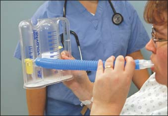
Auscultate the patient’s lungs to provide a baseline for comparison with posttreatment auscultation. Instruct the patient to exhale normally, insert the mouthpiece, close the lips tightly around it because a weak seal may alter flow or volume readings, and then inhale as slowly and as deeply as possible (as shown below). If the patient has difficulty with this step, suggest sucking as one would through a straw but more slowly. Ask the patient to retain the entire volume of inhaled air for 3 seconds or, if you’re using a device with a light indicator, until the light turns off. This deep breath creates sustained transpulmonary pressure near the end of inspiration and is sometimes called a sustained maximal inspiration.

Tell the patient to remove the mouthpiece and exhale normally. Let the patient relax and take several normal breaths before attempting another breath with the spirometer. Repeat this sequence 5 to 10 times during every waking hour. Note tidal volumes.
Evaluate the patient’s ability to cough effectively, encouraging coughing during and after each exercise session because deep lung inflation may loosen secretions and facilitate their removal. Observe any expectorated secretions.
Auscultate the patient’s lungs, and compare findings with the first auscultation.

Stay updated, free articles. Join our Telegram channel

Full access? Get Clinical Tree


Get Clinical Tree app for offline access
