Developmental dysplasia of the hip
Description
Abnormal formation of the hip joint present at birth
Most common disorder affecting the hip joints in children younger than age 3 years
Can be unilateral or bilateral
About 85% of affected infants are females
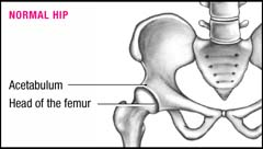
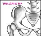
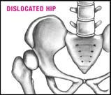
Occurs in three forms of varying severity (see Degrees of hip dysplasia)
Also called DDH
Pathophysiology
Normal fetal development of the hip joint requires that the femoral head be seated tightly within the acetabulum.
Any situation that disrupts this intimate relationship increases the risk of hip dysplasia, and may occur in utero, perinatally, or during infancy and childhood.
Displacement of the femoral head from within the acetabulum may damage joint structures, including articulating surfaces, blood vessels, tendons, ligaments, and nerves.
Disruption of blood flow to the joint may lead to ischemic necrosis of the femoral head.
Degrees of hip dysplasia
Normally, the head of the femur fits snugly into the acetabulum, allowing the hip to move properly. In congenital hip dysplasia, flattening of the acetabulum prevents the head of the femur from rotating adequately. The child’s hip may be unstable, subluxated (partially dislocated), or completely dislocated, with the femoral head lying totally outside the acetabulum. The degree of dysplasia and the child’s age are considered in determining the treatment choice.
 |
 |  |
Stay updated, free articles. Join our Telegram channel

Full access? Get Clinical Tree


Get Clinical Tree app for offline access
