Cirrhosis
Cirrhosis is a condition of chronic, progressive, irreversible damage to the liver resulting in compromised hepatic functioning due to a variety of causes. The seventh leading cause of death in the United States, it affects about 11 million Americans. Although caused by many different conditions, in the United States cirrhosis is most often caused by chronic alcohol consumption. In Third World and developing countries, it usually results from chronic hepatitis, either B, C, or D. Biliary cirrhosis is due to inflammation and scarring of the bile ducts and liver. There is a genetic component to this type of cirrhosis. Other types of cirrhosis include postnecrotic cirrhosis from hepatitis infection and metabolic cirrhosis from diseases such as Wilson’s disease, glycogen storage disease, or α1-antitrypsin deficiencies. Symptoms are similar regardless of the cause. Fifty percent with an established diagnosis of cirrhosis survive for 2 years; 35% survive 5 years. About 40% of cases are discovered on autopsy.
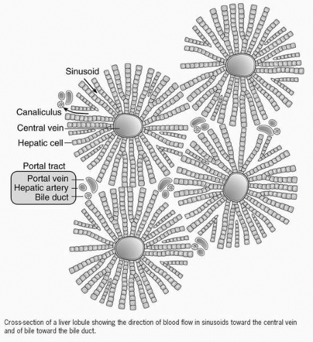 Figure 55-1 Cross-section of a liver lobule. |
The architecture of the liver is precisely arranged, with each cell and structure having a specific function to perform. If only small areas are affected, the liver can maintain vital functions, but as larger sections are destroyed, its abilities are taxed beyond repair. Damaged liver cells are replaced by tissue that is thick, rigid, inflexible, and incapable of performing any of the functions of healthy hepatocytes. This formation of scar tissue contracts, shrinking the organ and producing nodules in the hepatic parenchyma that is surrounded by fibrotic tissue with an irregular surface appearance that interferes with the normal vascular and bile pathways. Pressure gradients are affected, compromising blood flow in and out of the organ and backing up the bile. Bile stasis irritates and inflames hepatocytes, causing additional damage. Symptoms develop as the liver is first compromised and then fails.
 Biliary cirrhosis occurs due to inflammation and scarring of the bile ducts and liver.
 This formation of scar tissue contracts, shrinking the organ and producing nodules in the hepatic parenchyma that is surrounded by fibrotic tissue with an irregular surface appearance that interferes with the normal vascular and bile pathways.
Stay updated, free articles. Join our Telegram channel

Full access? Get Clinical Tree


Get Clinical Tree app for offline access
