Anatomy and Physiology of the Gastrointestinal System
The gastrointestinal (GI) tract includes the mouth, pharynx, esophagus, stomach, and small and large intestines. These structures, together with the accessory organs of digestion-which include the salivary glands, the pancreas, and the biliary system-comprise the GI system. This system provides nutrients to the body by moving, storing, and absorbing nutrients; secreting digestive juices; and digesting food. It is also referred to as the alimentary canal.
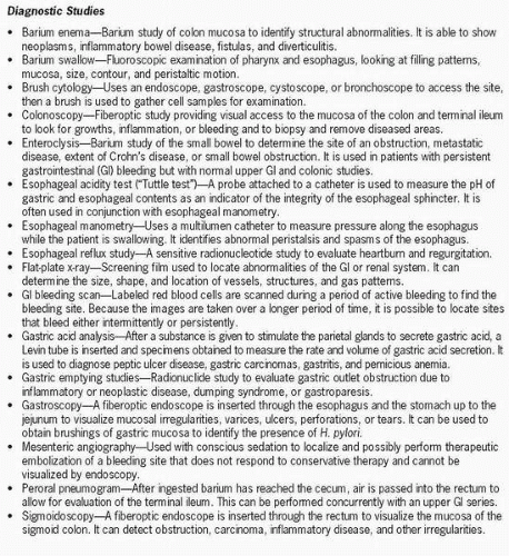 Figure 42-1 Diagnostic studies. |
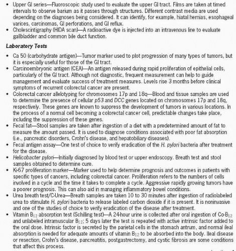 Figure 42-1 Continued. |
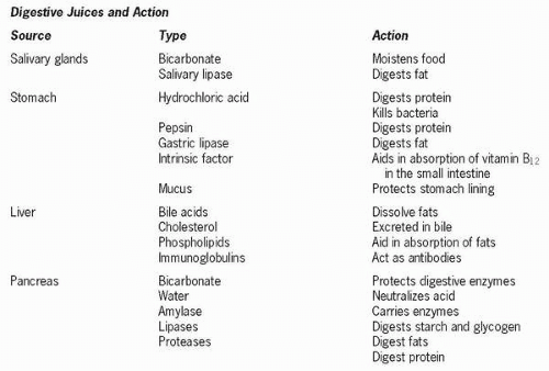 Figure 42-1 Continued. |
In the mouth, food is broken down and moistened by saliva secreted from the submandibular, parotid, and sublingual salivary glands. Food then becomes a semisolid mass and is swallowed. Reflex muscular movements propel the food bolus into the pharynx, through the laryngopharynx, and into the esophagus, which is a muscular tube about 25 cm long. Strong muscular contractions and mucus secreted from epithelial cells in the lining move the bolus along. The upper esophageal sphincter prevents the food bolus from entering the trachea or posterior aspect of the pharynx. At the lower end of the esophagus near its junction with the stomach is the lower esophageal sphincter, which serves as a barrier between the stomach and the esophagus. This site remains tonically constricted to prevent reflux of acidic gastric contents but relaxes during peristalsis, allowing passage of food into the stomach.
 The upper esophageal sphincter prevents the food bolus from entering the trachea or posterior aspect of the pharynx.
Once the bolus moves into the stomach, the reservoir that holds food, digestion continues. The stomach is divided into five anatomical areas: the cardia, which connects it to the esophagus; the fundus, the uppermost portion; the body, the largest section; the antrum, the lower section; and the pylorus, which connects the stomach to the duodenum. Here the pyloric sphincter, a muscular opening that controls gastric emptying, limits the reflux of bile from the small intestine back into the stomach. The gastric glands in the stomach contain specialized cells that act to either promote the digestive process or protect structures involved. These cells are chief cells, producing an inactivated form of the digestive enzyme pepsin, which converts proteins into proteoses and peptones; parietal cells, producing hydrochloric acid, an intrinsic factor needed for vitamin B12 absorption; mucous cells, producing an alkaline mucus that protects the stomach lining; and gastrin cells, which monitor pH. A 1-mm-thick layer of mucus protects the stomach lining from digestive juices.
Stay updated, free articles. Join our Telegram channel

Full access? Get Clinical Tree


