Anatomy and Physiology of the Cardiovascular System: Part II
The heart is a muscular organ located in the chest, between the lungs and above the diaphragm. Its four chambers are designed to work in a systematic fashion, each relying on the ability to work efficiently. There are several factors that influence the size and weight of the heart, including gender, age, and body weight. Disease states also affect the quality of heart function.
Regulation and coordination of contractions of the atria and ventricles are essential for effective pumping of the heart. There are both intrinsic and extrinsic mechanisms to accomplish these goals.
Intrinsic mechanisms achieve coordination through precise timing and routing of electrical impulse formation and conduction, which are
tied to stimulation of contractility. In the healthy heart, impulses originate in the sinoatrial (SA) node and spread rapidly through the atrial myocardial fibers that respond by contracting. These impulses are further conducted toward the atrioventricular (AV) node, where there is a slight delay before being transmitted to the ventricles. The impulse then continues through the His-Purkinje system and the ventricles and ultimately stimulates ventricular contraction.
tied to stimulation of contractility. In the healthy heart, impulses originate in the sinoatrial (SA) node and spread rapidly through the atrial myocardial fibers that respond by contracting. These impulses are further conducted toward the atrioventricular (AV) node, where there is a slight delay before being transmitted to the ventricles. The impulse then continues through the His-Purkinje system and the ventricles and ultimately stimulates ventricular contraction.
Four properties of cardiac tissue enable the initiation and conduction of impulses and myocardial contractility: automaticity, rhythmicity, conductivity, and excitability. Automaticity is the ability of specialized tissue cells in the SA node, parts of the atria, and AV node to spontaneously initiate impulses. Rhythmicity regulates the generation of impulses. Conductivity is the ability to transmit impulses from one fiber to another. Excitability is the ability to respond to an impulse and generate an action.
Cardiac cells achieve these tasks by initiating and conducting action potential (AP), which is a self-propagating wave of depolarization followed by repolarization. The APs are generated by sodium ions (Na+), potassium ions (K+), and calcium ions (Ca++) moving in and out of cells through channels in the cell membranes. In a resting myocardial cell, negatively charged ions line up inside the cell membrane, whereas positively charged ions line up outside, creating an electrical charge difference, and the cell is said to be polarized. With or without stimulation, channels in the membrane open, allowing positive ions to enter and eliminating the charge differences, and the cell is then said to be depolarized. After depolarization, the positively charged ions are extruded and the cell membrane repolarized.
Cardiac excitation normally begins in the SA node where autorhythmic fibers undergo rapid spontaneous depolarization, initiating APs at a rate of 90 to 100 times per minute, before any spontaneous depolarization in other regions. APs from the SA node spread to other areas of the conduction system, stimulating them before they are able to generate an AP at their own slower rate. Thus the SA node becomes the primary pacemaker for the heart. Hormones or neurotransmitters can slow or speed pacing of the heart. Slow conduction in the small size fibers of the AV node allows time for the atria to contract, moving blood forward into the ventricles before they contract.
If, because of disease or damage, the SA node fails to initiate an impulse, the slower AV node fibers can pick up the pacing chores; however,
with AV pacing, the pacing rate is 40 to 50 beats per minute. If activity in the nodes is suppressed, the heartbeat may be maintained by autorhythmic fibers in the ventricles such as the AV bundle, a bundle branch, or conduction myofibrils. These fibers fire AVs very slowly, only about 20 to 40 beats per minute, a rate that is too slow to adequately perfuse the brain. Sometimes a site other than the SA node develops abnormal self-excitability. Such a site is called an ectopic focus.
with AV pacing, the pacing rate is 40 to 50 beats per minute. If activity in the nodes is suppressed, the heartbeat may be maintained by autorhythmic fibers in the ventricles such as the AV bundle, a bundle branch, or conduction myofibrils. These fibers fire AVs very slowly, only about 20 to 40 beats per minute, a rate that is too slow to adequately perfuse the brain. Sometimes a site other than the SA node develops abnormal self-excitability. Such a site is called an ectopic focus.
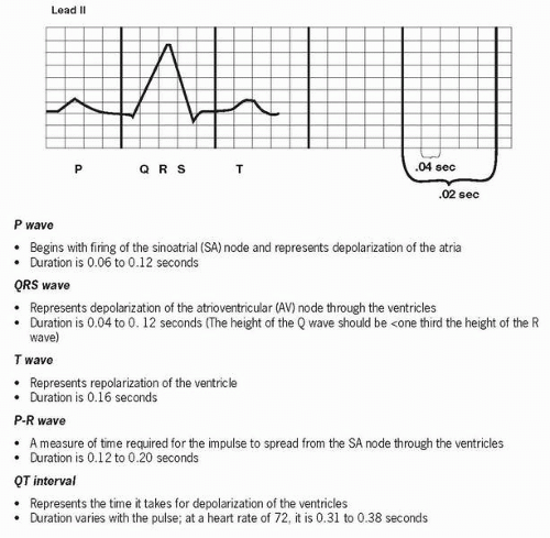
The level of excitability of myocardial cells is determined by the length of time since the previous depolarization. The recovery time is called the refractory period and is subdivided into the absolute refractory period, which extends through depolarization and most of repolarization when no other AP can be stimulated, and relative refractory period, which occurs slightly later when excitability is more likely (i.e., approximately on the second half of the T wave on an electrocardiogram [ECG]). The refractory period is normally longer than the period of contraction, giving the ventricles time to fill before another contraction can occur.
Stay updated, free articles. Join our Telegram channel

Full access? Get Clinical Tree


